 Movie
Movie Controller


+ Open data

- Basic information
| Entry | Database: PDB / ID: 7pee | |||||||||
|---|---|---|---|---|---|---|---|---|---|---|
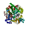
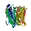
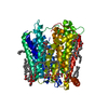
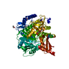
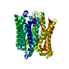
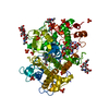
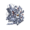
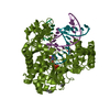
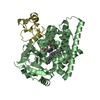
| Title | Crystal structure of extracellular part of human Trop2 | |||||||||
 Components Components | Tumor-associated calcium signal transducer 2 | |||||||||
 Keywords Keywords | MEMBRANE PROTEIN / extracellular part / dimer | |||||||||
| Function / homology |  Function and homology information Function and homology informationnegative regulation of branching involved in ureteric bud morphogenesis / ureteric bud morphogenesis / negative regulation of ruffle assembly / negative regulation of substrate adhesion-dependent cell spreading / negative regulation of epithelial cell migration / positive regulation of stem cell differentiation / negative regulation of stress fiber assembly / regulation of epithelial cell proliferation / negative regulation of cell motility / lateral plasma membrane ...negative regulation of branching involved in ureteric bud morphogenesis / ureteric bud morphogenesis / negative regulation of ruffle assembly / negative regulation of substrate adhesion-dependent cell spreading / negative regulation of epithelial cell migration / positive regulation of stem cell differentiation / negative regulation of stress fiber assembly / regulation of epithelial cell proliferation / negative regulation of cell motility / lateral plasma membrane / visual perception / basal plasma membrane / extracellular space / extracellular exosome / membrane / nucleus / cytosol Similarity search - Function | |||||||||
| Biological species |  Homo sapiens (human) Homo sapiens (human) | |||||||||
| Method |  X-RAY DIFFRACTION / SYNCHROTRON / MOLECULAR REPLACEMENT / Resolution: 2.81 Å X-RAY DIFFRACTION / SYNCHROTRON / MOLECULAR REPLACEMENT / Resolution: 2.81 Å | |||||||||
 Authors Authors | Pavsic, M. | |||||||||
| Funding support |  Slovenia, 2items Slovenia, 2items
| |||||||||
 Citation Citation | Journal: Int J Mol Sci / Year: 2021 Title: Trop2 Forms a Stable Dimer with Significant Structural Differences within the Membrane-Distal Region as Compared to EpCAM. Authors: Pavsic, M. | |||||||||
| History |
|
- Structure visualization
| Structure viewer | Molecule: MolmilJmol/JSmol |
|---|
- Downloads & links
-Download
| PDBx/mmCIF format | 7pee.cif.gz | 216.9 KB | Display | PDBx/mmCIF format |
|---|---|---|---|---|
| PDB format | pdb7pee.ent.gz | 159.3 KB | Display | PDB format |
| PDBx/mmJSON format | 7pee.json.gz | Tree view | PDBx/mmJSON format | |
| Others |  Other downloads Other downloads |
-Validation report
| Arichive directory | https://data.pdbj.org/pub/pdb/validation_reports/pe/7peeftp://data.pdbj.org/pub/pdb/validation_reports/pe/7pee | HTTPS FTP |
|---|
-Related structure data
| Related structure data |  4mzvS S: Starting model for refinement |
|---|---|
| Similar structure data |
-Links
 PDBj
PDBj- Assembly
| Deposited unit | 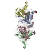
| ||||||||||||
|---|---|---|---|---|---|---|---|---|---|---|---|---|---|
| 1 | 
| ||||||||||||
| 2 | 
| ||||||||||||
| Unit cell |
|
-Components
| #1: Protein | Mass: 28381.301 Da / Num. of mol.: 4 / Mutation: N120Q,N208Q Source method: isolated from a genetically manipulated source Source: (gene. exp.) Homo sapiens (human) / Gene: TACSTD2, GA733-1, M1S1, TROP2 / Cell line (production host): Sf9 / Production host:   Spodoptera frugiperda (fall armyworm) / References: UniProt: P09758 Spodoptera frugiperda (fall armyworm) / References: UniProt: P09758#2: Polysaccharide | Source method: isolated from a genetically manipulated source #3: Chemical |   Mass: 96.063 Da / Num. of mol.: 2 / Source method: isolated from a natural source / Formula: SO4 Mass: 96.063 Da / Num. of mol.: 2 / Source method: isolated from a natural source / Formula: SO4#4: Sugar |   Type: D-saccharide, beta linking / Mass: 221.208 Da / Num. of mol.: 2 / Source method: isolated from a natural source / Formula: C8H15NO6 Type: D-saccharide, beta linking / Mass: 221.208 Da / Num. of mol.: 2 / Source method: isolated from a natural source / Formula: C8H15NO6#5: Water | ChemComp-HOH / |  Mass: 18.015 Da / Num. of mol.: 30 / Source method: isolated from a natural source / Formula: H2O Mass: 18.015 Da / Num. of mol.: 30 / Source method: isolated from a natural source / Formula: H2OHas ligand of interest | N | Has protein modification | Y | |
|---|
-Experimental details
-Experiment
| Experiment | Method: X-RAY DIFFRACTION / Number of used crystals: 1 |
|---|
- Sample preparation
| Crystal | Density Matthews: 5.05 Å3/Da / Density % sol: 75.63 % / Description: rod |
|---|---|
| Crystal grow | Temperature: 294 K / Method: vapor diffusion, sitting drop / pH: 5.8 Details: 1.6 M ammonium sulfate, 0.5 M NaCl, 0.0133 M EDTA, 0.1 M MES pH 5.8 |
-Data collection
| Diffraction | Mean temperature: 100 K / Serial crystal experiment: N |
|---|---|
| Diffraction source | Source: SYNCHROTRON / Site: ELETTRA  / Beamline: 11.2C / Wavelength: 0.9789 Å / Beamline: 11.2C / Wavelength: 0.9789 Å |
| Detector | Type: DECTRIS PILATUS 6M / Detector: PIXEL / Date: Feb 7, 2019 |
| Radiation | Protocol: SINGLE WAVELENGTH / Monochromatic (M) / Laue (L): M / Scattering type: x-ray |
| Radiation wavelength | Wavelength: 0.9789 Å / Relative weight: 1 |
| Reflection | Resolution: 2.81→48.38 Å / Num. obs: 57322 / % possible obs: 99.6 % / Redundancy: 6.6 % / Biso Wilson estimate: 64.21 Å2 / CC1/2: 0.998 / Rsym value: 0.1157 / Net I/σ(I): 15.14 |
| Reflection shell | Resolution: 2.81→2.98 Å / Mean I/σ(I) obs: 1.85 / Num. unique obs: 9084 / CC1/2: 0.717 / Rsym value: 1.19 / % possible all: 99.4 |
- Processing
| Software |
| |||||||||||||||||||||||||||||||||||||||||||||||||||||||||||||||||||||||||||||||||||||||||||||||||||||||||||||||||||||||||||||||||||||||||||||||||||
|---|---|---|---|---|---|---|---|---|---|---|---|---|---|---|---|---|---|---|---|---|---|---|---|---|---|---|---|---|---|---|---|---|---|---|---|---|---|---|---|---|---|---|---|---|---|---|---|---|---|---|---|---|---|---|---|---|---|---|---|---|---|---|---|---|---|---|---|---|---|---|---|---|---|---|---|---|---|---|---|---|---|---|---|---|---|---|---|---|---|---|---|---|---|---|---|---|---|---|---|---|---|---|---|---|---|---|---|---|---|---|---|---|---|---|---|---|---|---|---|---|---|---|---|---|---|---|---|---|---|---|---|---|---|---|---|---|---|---|---|---|---|---|---|---|---|---|---|---|
| Refinement | Method to determine structure: MOLECULAR REPLACEMENT Starting model: 4MZV Resolution: 2.81→48.37 Å / SU ML: 0.4202 / Cross valid method: FREE R-VALUE / σ(F): 1.34 / Phase error: 28.8396 Stereochemistry target values: GeoStd + Monomer Library + CDL v1.2
| |||||||||||||||||||||||||||||||||||||||||||||||||||||||||||||||||||||||||||||||||||||||||||||||||||||||||||||||||||||||||||||||||||||||||||||||||||
| Solvent computation | Shrinkage radii: 0.9 Å / VDW probe radii: 1.11 Å / Solvent model: FLAT BULK SOLVENT MODEL | |||||||||||||||||||||||||||||||||||||||||||||||||||||||||||||||||||||||||||||||||||||||||||||||||||||||||||||||||||||||||||||||||||||||||||||||||||
| Displacement parameters | Biso mean: 75.8 Å2 | |||||||||||||||||||||||||||||||||||||||||||||||||||||||||||||||||||||||||||||||||||||||||||||||||||||||||||||||||||||||||||||||||||||||||||||||||||
| Refinement step | Cycle: LAST / Resolution: 2.81→48.37 Å
| |||||||||||||||||||||||||||||||||||||||||||||||||||||||||||||||||||||||||||||||||||||||||||||||||||||||||||||||||||||||||||||||||||||||||||||||||||
| Refine LS restraints |
| |||||||||||||||||||||||||||||||||||||||||||||||||||||||||||||||||||||||||||||||||||||||||||||||||||||||||||||||||||||||||||||||||||||||||||||||||||
| LS refinement shell |
|
