 Movie
Movie Controller

[English] 日本語
 Yorodumi
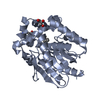
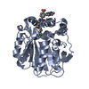
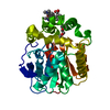
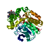
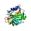
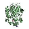
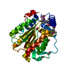
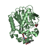
- PDB-7pcw: X-RAY STRUCTURE OF THE HALOALKANE DEHALOGENASE HALOTAG7-M175W LAB... -
+ Open data

- Basic information
| Entry | Database: PDB / ID: 7pcw | ||||||
|---|---|---|---|---|---|---|---|
| Title | X-RAY STRUCTURE OF THE HALOALKANE DEHALOGENASE HALOTAG7-M175W LABELED WITH A CHLOROALKANE-TETRAMETHYLRHODAMINE FLUOROPHORE SUBSTRATE | ||||||
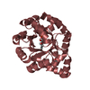
 Components Components | Haloalkane dehalogenase | ||||||
 Keywords Keywords | HYDROLASE / HALOALKANE DEHALOGENASE / HALO / TAG / HALOTAG7 / SELF-LABELING PROTEIN / FLUOROPHORE / TETRAMETHYLRHODAMINE | ||||||
| Function / homology |  Function and homology information Function and homology informationhaloalkane dehalogenase / haloalkane dehalogenase activity / response to toxic substance / membrane Similarity search - Function | ||||||
| Biological species |  Rhodococcus sp. (bacteria) Rhodococcus sp. (bacteria) | ||||||
| Method |  X-RAY DIFFRACTION / SYNCHROTRON / MOLECULAR REPLACEMENT / Resolution: 2.3 Å X-RAY DIFFRACTION / SYNCHROTRON / MOLECULAR REPLACEMENT / Resolution: 2.3 Å | ||||||
 Authors Authors | Tarnawski, M. / Frei, M. / Hiblot, J. / Johnsson, K. | ||||||
| Funding support | 1items
| ||||||
 Citation Citation | Journal: Nat.Methods / Year: 2022 Title: Engineered HaloTag variants for fluorescence lifetime multiplexing. Authors: Frei, M.S. / Tarnawski, M. / Roberti, M.J. / Koch, B. / Hiblot, J. / Johnsson, K. | ||||||
| History |
|
- Structure visualization
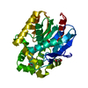
| Structure viewer | Molecule: MolmilJmol/JSmol |
|---|
- Downloads & links
-Download
| PDBx/mmCIF format | 7pcw.cif.gz | 301.3 KB | Display | PDBx/mmCIF format |
|---|---|---|---|---|
| PDB format | pdb7pcw.ent.gz | 195.8 KB | Display | PDB format |
| PDBx/mmJSON format | 7pcw.json.gz | Tree view | PDBx/mmJSON format | |
| Others |  Other downloads Other downloads |
-Validation report
| Arichive directory | https://data.pdbj.org/pub/pdb/validation_reports/pc/7pcwftp://data.pdbj.org/pub/pdb/validation_reports/pc/7pcw | HTTPS FTP |
|---|
-Related structure data
| Related structure data | 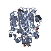 6zvyC 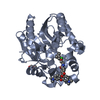 7pcxC 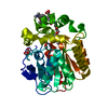 6y7aS C: citing same article ( S: Starting model for refinement |
|---|---|
| Similar structure data |


-Links
 PDBj
PDBj


- Assembly
| Deposited unit | 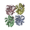
| ||||||||||||
|---|---|---|---|---|---|---|---|---|---|---|---|---|---|
| 1 | 
| ||||||||||||
| 2 | 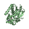
| ||||||||||||
| 3 | 
| ||||||||||||
| 4 | 
| ||||||||||||
| Unit cell |
|
-Components
| #1: Protein | Mass: 33280.996 Da / Num. of mol.: 4 Source method: isolated from a genetically manipulated source Source: (gene. exp.) Rhodococcus sp. (bacteria) / Gene: dhaA / Production host: #2: Chemical | ChemComp-OEH / [ 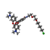  Mass: 637.185 Da / Num. of mol.: 4 / Source method: obtained synthetically / Formula: C35H43ClN3O6 / Feature type: SUBJECT OF INVESTIGATION Mass: 637.185 Da / Num. of mol.: 4 / Source method: obtained synthetically / Formula: C35H43ClN3O6 / Feature type: SUBJECT OF INVESTIGATION#3: Chemical | ChemComp-CL /   Mass: 35.453 Da / Num. of mol.: 4 / Source method: obtained synthetically / Formula: Cl Mass: 35.453 Da / Num. of mol.: 4 / Source method: obtained synthetically / Formula: Cl#4: Water | ChemComp-HOH / |  Mass: 18.015 Da / Num. of mol.: 125 / Source method: isolated from a natural source / Formula: H2O Mass: 18.015 Da / Num. of mol.: 125 / Source method: isolated from a natural source / Formula: H2OHas ligand of interest | Y | Has protein modification | Y | |
|---|
-Experimental details
-Experiment
| Experiment | Method: X-RAY DIFFRACTION / Number of used crystals: 1 |
|---|
- Sample preparation
| Crystal | Density Matthews: 1.98 Å3/Da / Density % sol: 37.92 % |
|---|---|
| Crystal grow | Temperature: 293 K / Method: vapor diffusion, hanging drop Details: 0.1 M MES pH 6.0, 1.0 M lithium chloride, 22% (m/v) PEG 6000 |
-Data collection
| Diffraction | Mean temperature: 100 K / Serial crystal experiment: N |
|---|---|
| Diffraction source | Source: SYNCHROTRON / Site: SLS  / Beamline: X10SA / Wavelength: 1 Å / Beamline: X10SA / Wavelength: 1 Å |
| Detector | Type: DECTRIS EIGER2 X 16M / Detector: PIXEL / Date: Jun 26, 2021 |
| Radiation | Monochromator: Si(111) / Protocol: SINGLE WAVELENGTH / Monochromatic (M) / Laue (L): M / Scattering type: x-ray |
| Radiation wavelength | Wavelength: 1 Å / Relative weight: 1 |
| Reflection | Resolution: 2.3→50 Å / Num. obs: 43477 / % possible obs: 95.6 % / Redundancy: 2.67 % / Biso Wilson estimate: 28.14 Å2 / CC1/2: 0.993 / Rmerge(I) obs: 0.098 / Net I/σ(I): 8.44 |
| Reflection shell | Resolution: 2.3→2.4 Å / Redundancy: 2.58 % / Rmerge(I) obs: 0.467 / Mean I/σ(I) obs: 2.8 / Num. unique obs: 5246 / CC1/2: 0.769 / % possible all: 96.1 |
- Processing
| Software |
| |||||||||||||||||||||||||||||||||||||||||||||||||||||||||||||||||||||||||||||||||||||||||||||||||||||||||||||||||||||||
|---|---|---|---|---|---|---|---|---|---|---|---|---|---|---|---|---|---|---|---|---|---|---|---|---|---|---|---|---|---|---|---|---|---|---|---|---|---|---|---|---|---|---|---|---|---|---|---|---|---|---|---|---|---|---|---|---|---|---|---|---|---|---|---|---|---|---|---|---|---|---|---|---|---|---|---|---|---|---|---|---|---|---|---|---|---|---|---|---|---|---|---|---|---|---|---|---|---|---|---|---|---|---|---|---|---|---|---|---|---|---|---|---|---|---|---|---|---|---|---|---|
| Refinement | Method to determine structure: MOLECULAR REPLACEMENT Starting model: 6Y7A Resolution: 2.3→40.36 Å / SU ML: 0.2773 / Cross valid method: FREE R-VALUE / σ(F): 1.98 / Phase error: 25.3785 Stereochemistry target values: GeoStd + Monomer Library + CDL v1.2
| |||||||||||||||||||||||||||||||||||||||||||||||||||||||||||||||||||||||||||||||||||||||||||||||||||||||||||||||||||||||
| Solvent computation | Shrinkage radii: 0.9 Å / VDW probe radii: 1.11 Å / Solvent model: FLAT BULK SOLVENT MODEL | |||||||||||||||||||||||||||||||||||||||||||||||||||||||||||||||||||||||||||||||||||||||||||||||||||||||||||||||||||||||
| Displacement parameters | Biso mean: 29.57 Å2 | |||||||||||||||||||||||||||||||||||||||||||||||||||||||||||||||||||||||||||||||||||||||||||||||||||||||||||||||||||||||
| Refinement step | Cycle: LAST / Resolution: 2.3→40.36 Å
| |||||||||||||||||||||||||||||||||||||||||||||||||||||||||||||||||||||||||||||||||||||||||||||||||||||||||||||||||||||||
| Refine LS restraints |
| |||||||||||||||||||||||||||||||||||||||||||||||||||||||||||||||||||||||||||||||||||||||||||||||||||||||||||||||||||||||
| LS refinement shell |
|
