 Movie
Movie Controller


+ Open data

- Basic information
| Entry | Database: PDB / ID: 7paa | ||||||
|---|---|---|---|---|---|---|---|
| Title | JC polyomavirus VP1 in complex with scFv 29B1 | ||||||
 Components Components |
| ||||||
 Keywords Keywords | VIRAL PROTEIN / antibody / polyomavirus / VP1 / neutralization | ||||||
| Function / homology |  Function and homology information Function and homology informationT=7 icosahedral viral capsid / clathrin-dependent endocytosis of virus by host cell / virion attachment to host cell / host cell nucleus / structural molecule activity Similarity search - Function | ||||||
| Biological species |  Homo sapiens (human) Homo sapiens (human) 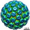 JC polyomavirus JC polyomavirus | ||||||
| Method |  X-RAY DIFFRACTION / SYNCHROTRON / MOLECULAR REPLACEMENT / Resolution: 3.102 Å X-RAY DIFFRACTION / SYNCHROTRON / MOLECULAR REPLACEMENT / Resolution: 3.102 Å | ||||||
 Authors Authors | Harprecht, C. / Stroeh, L.J. / Freytag, J. / Stehle, T. | ||||||
| Funding support |  Germany, 1items Germany, 1items
| ||||||
 Citation Citation | Journal: To Be Published Title: Structural characterization of human neutralizing antibodies against JC and BK polyomavirus Authors: Harprecht, C. / Stroeh, L.J. / Senn, L. / O'Hara, B.A. / Nagel, F. / Freytag, J. / Stiehrof, Y.D. / Atwood, W.J. / Combaluzier, B. / Stehle, T. | ||||||
| History |
|
- Structure visualization
| Structure viewer | Molecule: MolmilJmol/JSmol |
|---|
- Downloads & links
-Download
| PDBx/mmCIF format | 7paa.cif.gz | 928.9 KB | Display | PDBx/mmCIF format |
|---|---|---|---|---|
| PDB format | pdb7paa.ent.gz | Display | PDB format | |
| PDBx/mmJSON format | 7paa.json.gz | Tree view | PDBx/mmJSON format | |
| Others |  Other downloads Other downloads |
-Validation report
| Arichive directory | https://data.pdbj.org/pub/pdb/validation_reports/pa/7paaftp://data.pdbj.org/pub/pdb/validation_reports/pa/7paa | HTTPS FTP |
|---|
-Related structure data
| Related structure data |  7pa6C  7pa7C  7pa8C  7pa9C  3nxgS  4kq3S S: Starting model for refinement C: citing same article ( |
|---|---|
| Similar structure data |
-Links
 PDBj
PDBj


- Assembly
| Deposited unit | 
| ||||||||
|---|---|---|---|---|---|---|---|---|---|
| 1 | 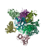
| ||||||||
| 2 | 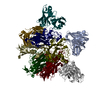
| ||||||||
| Unit cell |
|
-Components
| #1: Antibody | Mass: 26947.736 Da / Num. of mol.: 10 Source method: isolated from a genetically manipulated source Source: (gene. exp.) Homo sapiens (human) / Production host:  #2: Protein | Mass: 30075.861 Da / Num. of mol.: 10 / Fragment: UNP residues 23-290 Source method: isolated from a genetically manipulated source Source: (gene. exp.) JC polyomavirus / Production host: #3: Water | ChemComp-HOH / |  Mass: 18.015 Da / Num. of mol.: 23 / Source method: isolated from a natural source / Formula: H2O Mass: 18.015 Da / Num. of mol.: 23 / Source method: isolated from a natural source / Formula: H2OHas protein modification | Y | |
|---|
-Experimental details
-Experiment
| Experiment | Method: X-RAY DIFFRACTION / Number of used crystals: 1 |
|---|
- Sample preparation
| Crystal | Density Matthews: 2.9 Å3/Da / Density % sol: 57.63 % |
|---|---|
| Crystal grow | Temperature: 277.15 K / Method: vapor diffusion, sitting drop Details: 0.2 M trimethanolamine N-oxide, 0.1 M Tris (pH 8.5), 20% PEG 2000 |
-Data collection
| Diffraction | Mean temperature: 93 K / Serial crystal experiment: N |
|---|---|
| Diffraction source | Source: SYNCHROTRON / Site: SLS  / Beamline: X06DA / Wavelength: 0.9794 Å / Beamline: X06DA / Wavelength: 0.9794 Å |
| Detector | Type: DECTRIS PILATUS 2M-F / Detector: PIXEL / Date: Oct 24, 2016 |
| Radiation | Protocol: SINGLE WAVELENGTH / Monochromatic (M) / Laue (L): M / Scattering type: x-ray |
| Radiation wavelength | Wavelength: 0.9794 Å / Relative weight: 1 |
| Reflection | Resolution: 3→48.5 Å / Num. obs: 129715 / % possible obs: 99.8 % / Redundancy: 7.06 % / CC1/2: 0.99 / Rrim(I) all: 0.23 / Net I/σ(I): 10.37 |
| Reflection shell | Resolution: 3→3.18 Å / Mean I/σ(I) obs: 2.21 / Num. unique obs: 20801 / CC1/2: 0.72 / Rrim(I) all: 0.95 |
- Processing
| Software |
| |||||||||||||||||||||||||||||||||||||||||||||||||||||||||||||||||||||||||||||||||||||||||||||||||||||||||||||||||||||||||||||||||||||||||||||||||||||||||||||||||||||||||||||||||||||||||||||||||||||||||||||||||||||||||||||||||||||||
|---|---|---|---|---|---|---|---|---|---|---|---|---|---|---|---|---|---|---|---|---|---|---|---|---|---|---|---|---|---|---|---|---|---|---|---|---|---|---|---|---|---|---|---|---|---|---|---|---|---|---|---|---|---|---|---|---|---|---|---|---|---|---|---|---|---|---|---|---|---|---|---|---|---|---|---|---|---|---|---|---|---|---|---|---|---|---|---|---|---|---|---|---|---|---|---|---|---|---|---|---|---|---|---|---|---|---|---|---|---|---|---|---|---|---|---|---|---|---|---|---|---|---|---|---|---|---|---|---|---|---|---|---|---|---|---|---|---|---|---|---|---|---|---|---|---|---|---|---|---|---|---|---|---|---|---|---|---|---|---|---|---|---|---|---|---|---|---|---|---|---|---|---|---|---|---|---|---|---|---|---|---|---|---|---|---|---|---|---|---|---|---|---|---|---|---|---|---|---|---|---|---|---|---|---|---|---|---|---|---|---|---|---|---|---|---|---|---|---|---|---|---|---|---|---|---|---|---|---|---|---|---|---|
| Refinement | Method to determine structure: MOLECULAR REPLACEMENT Starting model: 3NXG, 4KQ3 Resolution: 3.102→48.507 Å / Cor.coef. Fo:Fc: 0.885 / Cor.coef. Fo:Fc free: 0.869 / WRfactor Rfree: 0.212 / WRfactor Rwork: 0.195 / Average fsc free: 0.9053 / Average fsc work: 0.9098 / Cross valid method: FREE R-VALUE / ESU R Free: 0.441 Details: Hydrogens have been added in their riding positions
| |||||||||||||||||||||||||||||||||||||||||||||||||||||||||||||||||||||||||||||||||||||||||||||||||||||||||||||||||||||||||||||||||||||||||||||||||||||||||||||||||||||||||||||||||||||||||||||||||||||||||||||||||||||||||||||||||||||||
| Solvent computation | Ion probe radii: 0.8 Å / Shrinkage radii: 0.8 Å / VDW probe radii: 1.2 Å / Solvent model: MASK BULK SOLVENT | |||||||||||||||||||||||||||||||||||||||||||||||||||||||||||||||||||||||||||||||||||||||||||||||||||||||||||||||||||||||||||||||||||||||||||||||||||||||||||||||||||||||||||||||||||||||||||||||||||||||||||||||||||||||||||||||||||||||
| Displacement parameters | Biso mean: 44.614 Å2
| |||||||||||||||||||||||||||||||||||||||||||||||||||||||||||||||||||||||||||||||||||||||||||||||||||||||||||||||||||||||||||||||||||||||||||||||||||||||||||||||||||||||||||||||||||||||||||||||||||||||||||||||||||||||||||||||||||||||
| Refinement step | Cycle: LAST / Resolution: 3.102→48.507 Å
| |||||||||||||||||||||||||||||||||||||||||||||||||||||||||||||||||||||||||||||||||||||||||||||||||||||||||||||||||||||||||||||||||||||||||||||||||||||||||||||||||||||||||||||||||||||||||||||||||||||||||||||||||||||||||||||||||||||||
| Refine LS restraints |
| |||||||||||||||||||||||||||||||||||||||||||||||||||||||||||||||||||||||||||||||||||||||||||||||||||||||||||||||||||||||||||||||||||||||||||||||||||||||||||||||||||||||||||||||||||||||||||||||||||||||||||||||||||||||||||||||||||||||
| LS refinement shell | Refine-ID: X-RAY DIFFRACTION / Total num. of bins used: 20
|
