 Movie
Movie Controller


+ Open data

- Basic information
| Entry | Database: PDB / ID: 7p9n | ||||||
|---|---|---|---|---|---|---|---|
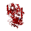
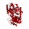
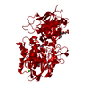
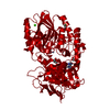
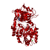
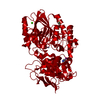
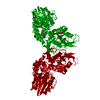
| Title | Crystal structure of CD73 in complex with AMP in the open form | ||||||
 Components Components | 5'-nucleotidase | ||||||
 Keywords Keywords | HYDROLASE / ecto-5'-nucleotidase / eN / substrate | ||||||
| Function / homology |  Function and homology information Function and homology informationthymidylate 5'-phosphatase / ADP catabolic process / 5'-deoxynucleotidase / 5'-deoxynucleotidase activity / 7-methylguanosine nucleotidase / adenosine biosynthetic process / inhibition of non-skeletal tissue mineralization / Pyrimidine catabolism / IMP-specific 5'-nucleotidase / AMP catabolic process ...thymidylate 5'-phosphatase / ADP catabolic process / 5'-deoxynucleotidase / 5'-deoxynucleotidase activity / 7-methylguanosine nucleotidase / adenosine biosynthetic process / inhibition of non-skeletal tissue mineralization / Pyrimidine catabolism / IMP-specific 5'-nucleotidase / AMP catabolic process / Purine catabolism / 5'-nucleotidase / 5'-nucleotidase activity / Nicotinate metabolism / leukocyte cell-cell adhesion / DNA metabolic process / response to ATP / Purinergic signaling in leishmaniasis infection / ATP metabolic process / calcium ion homeostasis / negative regulation of inflammatory response / external side of plasma membrane / nucleotide binding / cell surface / extracellular exosome / zinc ion binding / nucleoplasm / membrane / identical protein binding / plasma membrane / cytosol Similarity search - Function | ||||||
| Biological species |  Homo sapiens (human) Homo sapiens (human) | ||||||
| Method |  X-RAY DIFFRACTION / SYNCHROTRON / FOURIER SYNTHESIS / Resolution: 1.55 Å X-RAY DIFFRACTION / SYNCHROTRON / FOURIER SYNTHESIS / Resolution: 1.55 Å | ||||||
 Authors Authors | Scaletti, E.R. / Strater, N. | ||||||
 Citation Citation | Journal: Purinergic Signal / Year: 2021 Title: Substrate binding modes of purine and pyrimidine nucleotides to human ecto-5'-nucleotidase (CD73) and inhibition by their bisphosphonic acid derivatives. Authors: Scaletti, E. / Huschmann, F.U. / Mueller, U. / Weiss, M.S. / Strater, N. | ||||||
| History |
|
- Structure visualization
| Structure viewer | Molecule: MolmilJmol/JSmol |
|---|
- Downloads & links
-Download
| PDBx/mmCIF format | 7p9n.cif.gz | 231.2 KB | Display | PDBx/mmCIF format |
|---|---|---|---|---|
| PDB format | pdb7p9n.ent.gz | 180.5 KB | Display | PDB format |
| PDBx/mmJSON format | 7p9n.json.gz | Tree view | PDBx/mmJSON format | |
| Others |  Other downloads Other downloads |
-Validation report
| Arichive directory | https://data.pdbj.org/pub/pdb/validation_reports/p9/7p9nftp://data.pdbj.org/pub/pdb/validation_reports/p9/7p9n | HTTPS FTP |
|---|
-Related structure data
| Related structure data |  7p9rC  7p9tC  7pa4C  7pb5C 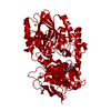 7pbaC  7pbbC  7pbyC  7pcpC  7pd9C  4h2gS S: Starting model for refinement C: citing same article ( |
|---|---|
| Similar structure data |


-Links
 PDBj
PDBj


- Assembly
| Deposited unit | 
| ||||||||
|---|---|---|---|---|---|---|---|---|---|
| 1 | 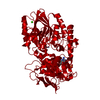
| ||||||||
| Unit cell |
| ||||||||
| Components on special symmetry positions |
|
-Components
-Protein , 1 types, 1 molecules A
| #1: Protein | Mass: 60464.340 Da / Num. of mol.: 1 / Mutation: T376A Source method: isolated from a genetically manipulated source Source: (gene. exp.) Homo sapiens (human) / Gene: NT5E, NT5, NTE / Production host:  |
|---|
-Non-polymers , 5 types, 475 molecules 


| #2: Chemical |  Mass: 65.409 Da / Num. of mol.: 2 / Source method: obtained synthetically / Formula: Zn Mass: 65.409 Da / Num. of mol.: 2 / Source method: obtained synthetically / Formula: Zn#3: Chemical | ChemComp-CA / |  Mass: 40.078 Da / Num. of mol.: 1 / Source method: obtained synthetically / Formula: Ca Mass: 40.078 Da / Num. of mol.: 1 / Source method: obtained synthetically / Formula: Ca#4: Chemical | ChemComp-GOL / |  Mass: 92.094 Da / Num. of mol.: 1 / Source method: obtained synthetically / Formula: C3H8O3 Mass: 92.094 Da / Num. of mol.: 1 / Source method: obtained synthetically / Formula: C3H8O3#5: Chemical | ChemComp-AMP / |  Mass: 347.221 Da / Num. of mol.: 1 / Source method: obtained synthetically / Formula: C10H14N5O7P / Feature type: SUBJECT OF INVESTIGATION / Comment: AMP*YM Mass: 347.221 Da / Num. of mol.: 1 / Source method: obtained synthetically / Formula: C10H14N5O7P / Feature type: SUBJECT OF INVESTIGATION / Comment: AMP*YM#6: Water | ChemComp-HOH / | Mass: 18.015 Da / Num. of mol.: 470 / Source method: isolated from a natural source / Formula: H2O |
|---|
-Details
| Has ligand of interest | Y |
|---|---|
| Has protein modification | Y |
-Experimental details
-Experiment
| Experiment | Method: X-RAY DIFFRACTION / Number of used crystals: 1 |
|---|
- Sample preparation
| Crystal | Density Matthews: 2.42 Å3/Da / Density % sol: 49.24 % |
|---|---|
| Crystal grow | Temperature: 292.15 K / Method: vapor diffusion, hanging drop / pH: 7.8 Details: 7 mg/mL protein concentration, 100 mM Tris pH 7.8, 10 % PEG6000, equal amounts of protein and reservoir. Following crystal formation (1-2 days), the crystals were transferred to soaking ...Details: 7 mg/mL protein concentration, 100 mM Tris pH 7.8, 10 % PEG6000, equal amounts of protein and reservoir. Following crystal formation (1-2 days), the crystals were transferred to soaking solution containing reservoir solution and 100 mM AMP. Crystals were then transferred to cryo solution containing an additional 20 % glycerol, soaked for ~2-5 min, and flash frozen in liquid nitrogen. |
-Data collection
| Diffraction | Mean temperature: 100 K / Serial crystal experiment: N | ||||||||||||||||||||||||||||||
|---|---|---|---|---|---|---|---|---|---|---|---|---|---|---|---|---|---|---|---|---|---|---|---|---|---|---|---|---|---|---|---|
| Diffraction source | Source: SYNCHROTRON / Site: BESSY  / Beamline: 14.1 / Wavelength: 0.91841 Å / Beamline: 14.1 / Wavelength: 0.91841 Å | ||||||||||||||||||||||||||||||
| Detector | Type: DECTRIS PILATUS 6M / Detector: PIXEL / Date: Jun 23, 2016 | ||||||||||||||||||||||||||||||
| Radiation | Protocol: SINGLE WAVELENGTH / Monochromatic (M) / Laue (L): M / Scattering type: x-ray | ||||||||||||||||||||||||||||||
| Radiation wavelength | Wavelength: 0.91841 Å / Relative weight: 1 | ||||||||||||||||||||||||||||||
| Reflection | Resolution: 1.55→47.27 Å / Num. obs: 78906 / % possible obs: 92.6 % / Redundancy: 6.2 % / CC1/2: 0.997 / Rmerge(I) obs: 0.103 / Rpim(I) all: 0.044 / Rrim(I) all: 0.113 / Net I/σ(I): 11.5 | ||||||||||||||||||||||||||||||
| Reflection shell | Diffraction-ID: 1
|
- Processing
| Software |
| ||||||||||||||||||||||||||||||||||||||||||||||||||||||||||||
|---|---|---|---|---|---|---|---|---|---|---|---|---|---|---|---|---|---|---|---|---|---|---|---|---|---|---|---|---|---|---|---|---|---|---|---|---|---|---|---|---|---|---|---|---|---|---|---|---|---|---|---|---|---|---|---|---|---|---|---|---|---|
| Refinement | Method to determine structure: FOURIER SYNTHESIS Starting model: 4h2g Resolution: 1.55→47.27 Å / Cor.coef. Fo:Fc: 0.964 / Cor.coef. Fo:Fc free: 0.953 / SU B: 2.662 / SU ML: 0.043 / Cross valid method: THROUGHOUT / σ(F): 0 / ESU R: 0.071 / ESU R Free: 0.072 / Stereochemistry target values: MAXIMUM LIKELIHOOD Details: HYDROGENS HAVE BEEN ADDED IN THE RIDING POSITIONS U VALUES : WITH TLS ADDED
| ||||||||||||||||||||||||||||||||||||||||||||||||||||||||||||
| Solvent computation | Ion probe radii: 0.8 Å / Shrinkage radii: 0.8 Å / VDW probe radii: 1.2 Å / Solvent model: MASK | ||||||||||||||||||||||||||||||||||||||||||||||||||||||||||||
| Displacement parameters | Biso max: 100.75 Å2 / Biso mean: 11.648 Å2 / Biso min: 5.42 Å2
| ||||||||||||||||||||||||||||||||||||||||||||||||||||||||||||
| Refinement step | Cycle: final / Resolution: 1.55→47.27 Å
| ||||||||||||||||||||||||||||||||||||||||||||||||||||||||||||
| Refine LS restraints |
| ||||||||||||||||||||||||||||||||||||||||||||||||||||||||||||
| LS refinement shell | Resolution: 1.554→1.594 Å / Rfactor Rfree error: 0 / Total num. of bins used: 20
| ||||||||||||||||||||||||||||||||||||||||||||||||||||||||||||
| Refinement TLS params. | Method: refined / Origin x: 24.3085 Å / Origin y: 24.361 Å / Origin z: 22.8304 Å
|
