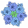
[English] 日本語
 Yorodumi
- PDB-7p7o: X-RAY CRYSTAL STRUCTURE OF SPOROSARCINA PASTEURII UREASE INHIBITE... -
+ Open data
- Basic information
| Entry | Database: PDB / ID: 7p7o | ||||||
|---|---|---|---|---|---|---|---|
| Title | X-RAY CRYSTAL STRUCTURE OF SPOROSARCINA PASTEURII UREASE INHIBITED BY THE GOLD(I)-DIPHOSPHINE COMPOUND Au(PEt3)2Cl DETERMINED AT 1.87 ANGSTROMS | ||||||
 Components Components | (Urease subunit ...) x 3 | ||||||
 Keywords Keywords | HYDROLASE / urease / nickel / gold / enzyme / urea | ||||||
| Function / homology |  Function and homology information Function and homology informationurease complex / urease / urease activity / urea catabolic process / nickel cation binding / cytoplasm Similarity search - Function | ||||||
| Biological species |  Sporosarcina pasteurii (bacteria) Sporosarcina pasteurii (bacteria) | ||||||
| Method |  X-RAY DIFFRACTION / X-RAY DIFFRACTION /  SYNCHROTRON / SYNCHROTRON /  MOLECULAR REPLACEMENT / Resolution: 1.87 Å MOLECULAR REPLACEMENT / Resolution: 1.87 Å | ||||||
 Authors Authors | Mazzei, L. / Ciurli, S. / Cianci, M. / Messori, L. / Massai, L. | ||||||
| Funding support | 1items
| ||||||
 Citation Citation |  Journal: Dalton Trans / Year: 2021 Journal: Dalton Trans / Year: 2021Title: Medicinal Au(I) compounds targeting urease as prospective antimicrobial agents: unveiling the structural basis for enzyme inhibition. Authors: Mazzei, L. / Massai, L. / Cianci, M. / Messori, L. / Ciurli, S. | ||||||
| History |
|
- Structure visualization
| Structure viewer | Molecule:  Molmil Molmil Jmol/JSmol Jmol/JSmol |
|---|
- Downloads & links
- Download
| PDBx/mmCIF format |  7p7o.cif.gz 7p7o.cif.gz | 194.3 KB | Display |  PDBx/mmCIF format PDBx/mmCIF format |
|---|---|---|---|---|
| PDB format |  pdb7p7o.ent.gz pdb7p7o.ent.gz | Display |  PDB format PDB format | |
| PDBx/mmJSON format |  7p7o.json.gz 7p7o.json.gz | Tree view |  PDBx/mmJSON format PDBx/mmJSON format | |
| Others |  Other downloads Other downloads |
-Validation report
| Summary document |  7p7o_validation.pdf.gz 7p7o_validation.pdf.gz | 1.3 MB | Display |  wwPDB validaton report wwPDB validaton report |
|---|---|---|---|---|
| Full document |  7p7o_full_validation.pdf.gz 7p7o_full_validation.pdf.gz | 1.3 MB | Display | |
| Data in XML |  7p7o_validation.xml.gz 7p7o_validation.xml.gz | 35.7 KB | Display | |
| Data in CIF |  7p7o_validation.cif.gz 7p7o_validation.cif.gz | 52.6 KB | Display | |
| Arichive directory |  https://data.pdbj.org/pub/pdb/validation_reports/p7/7p7o https://data.pdbj.org/pub/pdb/validation_reports/p7/7p7o ftp://data.pdbj.org/pub/pdb/validation_reports/p7/7p7o ftp://data.pdbj.org/pub/pdb/validation_reports/p7/7p7o | HTTPS FTP |
-Related structure data
| Related structure data |  7p7nC  5ol4S S: Starting model for refinement C: citing same article ( |
|---|---|
| Similar structure data | Similarity search - Function & homology  F&H Search F&H Search |
- Links
Links
- Assembly
| Deposited unit | 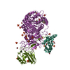
| ||||||||||||||||||
|---|---|---|---|---|---|---|---|---|---|---|---|---|---|---|---|---|---|---|---|
| 1 | 
| ||||||||||||||||||
| Unit cell |
| ||||||||||||||||||
| Components on special symmetry positions |
|
- Components
-Urease subunit ... , 3 types, 3 molecules AAABBBCCC
| #1: Protein | Mass: 11134.895 Da / Num. of mol.: 1 / Source method: isolated from a natural source / Source: (natural)  Sporosarcina pasteurii (bacteria) / References: UniProt: P41022, urease Sporosarcina pasteurii (bacteria) / References: UniProt: P41022, urease |
|---|---|
| #2: Protein | Mass: 13529.061 Da / Num. of mol.: 1 / Source method: isolated from a natural source / Source: (natural)  Sporosarcina pasteurii (bacteria) / References: UniProt: P41021, urease Sporosarcina pasteurii (bacteria) / References: UniProt: P41021, urease |
| #3: Protein | Mass: 61575.648 Da / Num. of mol.: 1 / Source method: isolated from a natural source / Source: (natural)  Sporosarcina pasteurii (bacteria) / References: UniProt: P41020, urease Sporosarcina pasteurii (bacteria) / References: UniProt: P41020, urease |
-Non-polymers , 6 types, 501 molecules 





| #4: Chemical | ChemComp-EDO / #5: Chemical | ChemComp-SO4 / #6: Chemical | #7: Chemical | ChemComp-O / | #8: Chemical | #9: Water | ChemComp-HOH / | |
|---|
-Details
| Has ligand of interest | N |
|---|
-Experimental details
-Experiment
| Experiment | Method:  X-RAY DIFFRACTION / Number of used crystals: 1 X-RAY DIFFRACTION / Number of used crystals: 1 |
|---|
- Sample preparation
| Crystal | Density Matthews: 2.76 Å3/Da / Density % sol: 55.6 % / Description: rice shaped |
|---|---|
| Crystal grow | Temperature: 293 K / Method: vapor diffusion, hanging drop / pH: 6.5 Details: THE PROTEIN-LIGAND (1.2 mM) COMPLEX IN 50 MM HEPES BUFFER, PH 7.50 (ALSO CONTAINING 10% (V/V) DMSO), DILUTED 1:1 WITH A SOLUTION OF 1.2-1.7 M AMMONIUM SULFATE ALSO CONTAINING THE SAME ...Details: THE PROTEIN-LIGAND (1.2 mM) COMPLEX IN 50 MM HEPES BUFFER, PH 7.50 (ALSO CONTAINING 10% (V/V) DMSO), DILUTED 1:1 WITH A SOLUTION OF 1.2-1.7 M AMMONIUM SULFATE ALSO CONTAINING THE SAME CONCENTRATION OF LIGAND AND DMSO. |
-Data collection
| Diffraction | Mean temperature: 100 K / Serial crystal experiment: N |
|---|---|
| Diffraction source | Source:  SYNCHROTRON / Site: SYNCHROTRON / Site:  PETRA III, EMBL c/o DESY PETRA III, EMBL c/o DESY  / Beamline: P13 (MX1) / Wavelength: 0.9762 Å / Beamline: P13 (MX1) / Wavelength: 0.9762 Å |
| Detector | Type: DECTRIS PILATUS 6M-F / Detector: PIXEL / Date: Nov 30, 2019 |
| Radiation | Monochromator: Si(111) silicon crystal / Protocol: SINGLE WAVELENGTH / Monochromatic (M) / Laue (L): M / Scattering type: x-ray |
| Radiation wavelength | Wavelength: 0.9762 Å / Relative weight: 1 |
| Reflection | Resolution: 1.87→114 Å / Num. obs: 80498 / % possible obs: 100 % / Redundancy: 17.4 % / Biso Wilson estimate: 26.89 Å2 / CC1/2: 0.999 / Rmerge(I) obs: 0.151 / Rpim(I) all: 0.038 / Rrim(I) all: 0.159 / Net I/σ(I): 17.9 |
| Reflection shell | Resolution: 1.87→1.91 Å / Redundancy: 17.5 % / Rmerge(I) obs: 3.098 / Mean I/σ(I) obs: 1.5 / Num. unique obs: 4557 / CC1/2: 0.744 / Rpim(I) all: 0.778 / Rrim(I) all: 3.296 / % possible all: 100 |
- Processing
| Software |
| |||||||||||||||||||||||||||||||||||||||||||||||||||||||||||||||||||||||||||||||||||||||||||||||||||||||||||||||||||||||||||||||||||||||||||||||||||
|---|---|---|---|---|---|---|---|---|---|---|---|---|---|---|---|---|---|---|---|---|---|---|---|---|---|---|---|---|---|---|---|---|---|---|---|---|---|---|---|---|---|---|---|---|---|---|---|---|---|---|---|---|---|---|---|---|---|---|---|---|---|---|---|---|---|---|---|---|---|---|---|---|---|---|---|---|---|---|---|---|---|---|---|---|---|---|---|---|---|---|---|---|---|---|---|---|---|---|---|---|---|---|---|---|---|---|---|---|---|---|---|---|---|---|---|---|---|---|---|---|---|---|---|---|---|---|---|---|---|---|---|---|---|---|---|---|---|---|---|---|---|---|---|---|---|---|---|---|
| Refinement | Method to determine structure:  MOLECULAR REPLACEMENT MOLECULAR REPLACEMENTStarting model: 5ol4 Resolution: 1.87→65.985 Å / Cor.coef. Fo:Fc: 0.975 / Cor.coef. Fo:Fc free: 0.956 / WRfactor Rfree: 0.178 / WRfactor Rwork: 0.137 / SU B: 3.947 / SU ML: 0.107 / Average fsc free: 0.8765 / Average fsc work: 0.8915 / Cross valid method: FREE R-VALUE / ESU R: 0.118 / ESU R Free: 0.12 Details: Hydrogens have been added in their riding positions
| |||||||||||||||||||||||||||||||||||||||||||||||||||||||||||||||||||||||||||||||||||||||||||||||||||||||||||||||||||||||||||||||||||||||||||||||||||
| Solvent computation | Ion probe radii: 0.8 Å / Shrinkage radii: 0.8 Å / VDW probe radii: 1.2 Å / Solvent model: MASK BULK SOLVENT | |||||||||||||||||||||||||||||||||||||||||||||||||||||||||||||||||||||||||||||||||||||||||||||||||||||||||||||||||||||||||||||||||||||||||||||||||||
| Displacement parameters | Biso mean: 38.018 Å2
| |||||||||||||||||||||||||||||||||||||||||||||||||||||||||||||||||||||||||||||||||||||||||||||||||||||||||||||||||||||||||||||||||||||||||||||||||||
| Refinement step | Cycle: LAST / Resolution: 1.87→65.985 Å
| |||||||||||||||||||||||||||||||||||||||||||||||||||||||||||||||||||||||||||||||||||||||||||||||||||||||||||||||||||||||||||||||||||||||||||||||||||
| Refine LS restraints |
| |||||||||||||||||||||||||||||||||||||||||||||||||||||||||||||||||||||||||||||||||||||||||||||||||||||||||||||||||||||||||||||||||||||||||||||||||||
| LS refinement shell |
|
 Movie
Movie Controller


 PDBj
PDBj