 Movie
Movie Controller

[English] 日本語
 Yorodumi
- PDB-7p71: The PDZ domain of MAGI1_2 complexed with the PDZ-binding motif of... -
+ Open data

- Basic information
| Entry | Database: PDB / ID: 7p71 | ||||||
|---|---|---|---|---|---|---|---|
| Title | The PDZ domain of MAGI1_2 complexed with the PDZ-binding motif of HPV35-E6 | ||||||
 Components Components |
| ||||||
 Keywords Keywords | PEPTIDE BINDING PROTEIN / PDZ / complex | ||||||
| Function / homology |  Function and homology information Function and homology informationAnxA2-p11 complex / membrane raft assembly / positive regulation of receptor-mediated endocytosis involved in cholesterol transport / positive regulation of vacuole organization / phospholipase A2 inhibitor activity / positive regulation of low-density lipoprotein particle clearance / positive regulation of vesicle fusion / symbiont-mediated perturbation of host apoptosis / myelin sheath adaxonal region / negative regulation of low-density lipoprotein particle receptor catabolic process ...AnxA2-p11 complex / membrane raft assembly / positive regulation of receptor-mediated endocytosis involved in cholesterol transport / positive regulation of vacuole organization / phospholipase A2 inhibitor activity / positive regulation of low-density lipoprotein particle clearance / positive regulation of vesicle fusion / symbiont-mediated perturbation of host apoptosis / myelin sheath adaxonal region / negative regulation of low-density lipoprotein particle receptor catabolic process / positive regulation of plasma membrane repair / positive regulation of plasminogen activation / PCSK9-AnxA2 complex / positive regulation of cell-cell adhesion / cadherin binding involved in cell-cell adhesion / cornified envelope / endothelial cell morphogenesis / Schmidt-Lanterman incisure / vesicle budding from membrane / calcium-dependent phospholipid binding / negative regulation of receptor internalization / plasma membrane protein complex / osteoclast development / Dissolution of Fibrin Clot / S100 protein binding / collagen fibril organization / vesicle membrane / epithelial cell apoptotic process / phosphatidylserine binding / alpha-actinin binding / positive regulation of receptor recycling / basement membrane / positive regulation of exocytosis / Smooth Muscle Contraction / bicellular tight junction / regulation of neurogenesis / fibrinolysis / cytoskeletal protein binding / phosphatidylinositol-4,5-bisphosphate binding / lipid droplet / Gene and protein expression by JAK-STAT signaling after Interleukin-12 stimulation / lung development / Turbulent (oscillatory, disturbed) flow shear stress activates signaling by PIEZO1 and integrins in endothelial cells / cell projection / cell-matrix adhesion / response to activity / cell periphery / PDZ domain binding / adherens junction / serine-type endopeptidase inhibitor activity / sarcolemma / mRNA transcription by RNA polymerase II / RNA polymerase II transcription regulator complex / : / nuclear matrix / calcium-dependent protein binding / cell-cell junction / azurophil granule lumen / cell junction / melanosome / late endosome membrane / protein-containing complex assembly / protease binding / angiogenesis / midbody / basolateral plasma membrane / symbiont-mediated suppression of host cytoplasmic pattern recognition receptor signaling pathway via inhibition of IRF3 activity / vesicle / symbiont-mediated perturbation of host ubiquitin-like protein modification / host cell cytoplasm / early endosome / cell surface receptor signaling pathway / cell adhesion / endosome / symbiont-mediated suppression of host type I interferon-mediated signaling pathway / lysosomal membrane / calcium ion binding / Neutrophil degranulation / DNA-templated transcription / regulation of DNA-templated transcription / nucleolus / host cell nucleus / cell surface / signal transduction / positive regulation of transcription by RNA polymerase II / extracellular space / DNA binding / RNA binding / extracellular exosome / extracellular region / zinc ion binding / nucleoplasm / ATP binding / identical protein binding / membrane / nucleus / plasma membrane / cytoplasm / cytosol Similarity search - Function | ||||||
| Biological species |  Homo sapiens (human) Homo sapiens (human) Human papillomavirus 35 Human papillomavirus 35 | ||||||
| Method |  X-RAY DIFFRACTION / SYNCHROTRON / MOLECULAR REPLACEMENT / Resolution: 2.6 Å X-RAY DIFFRACTION / SYNCHROTRON / MOLECULAR REPLACEMENT / Resolution: 2.6 Å | ||||||
 Authors Authors | Gogl, G. / Cousido-Siah, A. / Trave, G. | ||||||
 Citation Citation | Journal: Nat Commun / Year: 2022 Title: Quantitative fragmentomics allow affinity mapping of interactomes. Authors: Gogl, G. / Zambo, B. / Kostmann, C. / Cousido-Siah, A. / Morlet, B. / Durbesson, F. / Negroni, L. / Eberling, P. / Jane, P. / Nomine, Y. / Zeke, A. / Ostergaard, S. / Monsellier, E. / Vincentelli, R. / Trave, G. | ||||||
| History |
|
- Structure visualization
| Structure viewer | Molecule: MolmilJmol/JSmol |
|---|
- Downloads & links
-Download
| PDBx/mmCIF format | 7p71.cif.gz | 350.6 KB | Display | PDBx/mmCIF format |
|---|---|---|---|---|
| PDB format | pdb7p71.ent.gz | 290.1 KB | Display | PDB format |
| PDBx/mmJSON format | 7p71.json.gz | Tree view | PDBx/mmJSON format | |
| Others |  Other downloads Other downloads |
-Validation report
| Arichive directory | https://data.pdbj.org/pub/pdb/validation_reports/p7/7p71ftp://data.pdbj.org/pub/pdb/validation_reports/p7/7p71 | HTTPS FTP |
|---|
-Related structure data
| Related structure data |  7p70C  7p72C 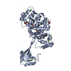 7p73C  7p74C  5n7dS S: Starting model for refinement C: citing same article ( |
|---|---|
| Similar structure data |
-Links
 PDBj
PDBj


- Assembly
| Deposited unit | 
| ||||||||
|---|---|---|---|---|---|---|---|---|---|
| 1 | 
| ||||||||
| 2 | 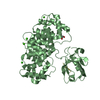
| ||||||||
| Unit cell |
|
-Components
-Protein / Protein/peptide , 2 types, 4 molecules ABCD
| #1: Protein | Mass: 48099.840 Da / Num. of mol.: 2 Source method: isolated from a genetically manipulated source Source: (gene. exp.) Homo sapiens (human)Gene: MAGI1, AIP3, BAIAP1, BAP1, TNRC19, ANXA2, ANX2, ANX2L4, CAL1H, LPC2D Production host:  #2: Protein/peptide | Mass: 1536.621 Da / Num. of mol.: 2 / Source method: obtained synthetically / Details: N-terminal biotin-ttds label / Source: (synth.) Human papillomavirus 35 / References: UniProt: P27228 |
|---|
-Non-polymers , 4 types, 29 molecules 


| #3: Chemical | ChemComp-CA /  Mass: 40.078 Da / Num. of mol.: 8 / Source method: obtained synthetically / Formula: Ca Mass: 40.078 Da / Num. of mol.: 8 / Source method: obtained synthetically / Formula: Ca#4: Chemical | ChemComp-GOL /  Mass: 92.094 Da / Num. of mol.: 5 / Source method: obtained synthetically / Formula: C3H8O3 Mass: 92.094 Da / Num. of mol.: 5 / Source method: obtained synthetically / Formula: C3H8O3#5: Chemical | ChemComp-CIT / |  Mass: 192.124 Da / Num. of mol.: 1 / Source method: obtained synthetically / Formula: C6H8O7 Mass: 192.124 Da / Num. of mol.: 1 / Source method: obtained synthetically / Formula: C6H8O7#6: Water | ChemComp-HOH / | Mass: 18.015 Da / Num. of mol.: 15 / Source method: isolated from a natural source / Formula: H2O |
|---|
-Details
| Has ligand of interest | N |
|---|
-Experimental details
-Experiment
| Experiment | Method: X-RAY DIFFRACTION / Number of used crystals: 1 |
|---|
- Sample preparation
| Crystal | Density Matthews: 2.92 Å3/Da / Density % sol: 57.86 % |
|---|---|
| Crystal grow | Temperature: 293 K / Method: vapor diffusion, hanging drop Details: 20-25% polyethylene glycol 3000, 100 mM sodium citrate buffered at pH 5.5 and 100 mM trisodium-citrate |
-Data collection
| Diffraction | Mean temperature: 100 K / Serial crystal experiment: N |
|---|---|
| Diffraction source | Source: SYNCHROTRON / Site: SLS  / Beamline: X06DA / Wavelength: 1 Å / Beamline: X06DA / Wavelength: 1 Å |
| Detector | Type: DECTRIS PILATUS 2M / Detector: PIXEL / Date: Jul 4, 2019 |
| Radiation | Protocol: SINGLE WAVELENGTH / Monochromatic (M) / Laue (L): M / Scattering type: x-ray |
| Radiation wavelength | Wavelength: 1 Å / Relative weight: 1 |
| Reflection | Resolution: 2.6→48 Å / Num. obs: 33417 / % possible obs: 94.4 % / Redundancy: 4.5 % / CC1/2: 1 / Rrim(I) all: 0.05 / Net I/σ(I): 19.06 |
| Reflection shell | Resolution: 2.6→2.67 Å / Mean I/σ(I) obs: 1.69 / Num. unique obs: 2471 / CC1/2: 0.852 / Rrim(I) all: 1.11 / % possible all: 94.5 |
- Processing
| Software |
| |||||||||||||||||||||||||||||||||||||||||||||||||||||||||||||||||||||||||||||||||||||||||||||||||||||||||||||||||||||||||||||||||||||||||||||||||||||||||||||||||||||||||||||||
|---|---|---|---|---|---|---|---|---|---|---|---|---|---|---|---|---|---|---|---|---|---|---|---|---|---|---|---|---|---|---|---|---|---|---|---|---|---|---|---|---|---|---|---|---|---|---|---|---|---|---|---|---|---|---|---|---|---|---|---|---|---|---|---|---|---|---|---|---|---|---|---|---|---|---|---|---|---|---|---|---|---|---|---|---|---|---|---|---|---|---|---|---|---|---|---|---|---|---|---|---|---|---|---|---|---|---|---|---|---|---|---|---|---|---|---|---|---|---|---|---|---|---|---|---|---|---|---|---|---|---|---|---|---|---|---|---|---|---|---|---|---|---|---|---|---|---|---|---|---|---|---|---|---|---|---|---|---|---|---|---|---|---|---|---|---|---|---|---|---|---|---|---|---|---|---|---|
| Refinement | Method to determine structure: MOLECULAR REPLACEMENT Starting model: 5N7D Resolution: 2.6→47.64 Å / SU ML: 0.58 / Cross valid method: THROUGHOUT / σ(F): 1.33 / Phase error: 37.89 / Stereochemistry target values: ML
| |||||||||||||||||||||||||||||||||||||||||||||||||||||||||||||||||||||||||||||||||||||||||||||||||||||||||||||||||||||||||||||||||||||||||||||||||||||||||||||||||||||||||||||||
| Solvent computation | Shrinkage radii: 1 Å / VDW probe radii: 1.2 Å / Solvent model: FLAT BULK SOLVENT MODEL | |||||||||||||||||||||||||||||||||||||||||||||||||||||||||||||||||||||||||||||||||||||||||||||||||||||||||||||||||||||||||||||||||||||||||||||||||||||||||||||||||||||||||||||||
| Displacement parameters | Biso max: 254.8 Å2 / Biso mean: 103.2257 Å2 / Biso min: 47.29 Å2 | |||||||||||||||||||||||||||||||||||||||||||||||||||||||||||||||||||||||||||||||||||||||||||||||||||||||||||||||||||||||||||||||||||||||||||||||||||||||||||||||||||||||||||||||
| Refinement step | Cycle: final / Resolution: 2.6→47.64 Å
| |||||||||||||||||||||||||||||||||||||||||||||||||||||||||||||||||||||||||||||||||||||||||||||||||||||||||||||||||||||||||||||||||||||||||||||||||||||||||||||||||||||||||||||||
| LS refinement shell | Refine-ID: X-RAY DIFFRACTION / Rfactor Rfree error: 0
| |||||||||||||||||||||||||||||||||||||||||||||||||||||||||||||||||||||||||||||||||||||||||||||||||||||||||||||||||||||||||||||||||||||||||||||||||||||||||||||||||||||||||||||||
| Refinement TLS params. | Method: refined / Refine-ID: X-RAY DIFFRACTION
| |||||||||||||||||||||||||||||||||||||||||||||||||||||||||||||||||||||||||||||||||||||||||||||||||||||||||||||||||||||||||||||||||||||||||||||||||||||||||||||||||||||||||||||||
| Refinement TLS group |
|
