 Movie
Movie Controller

[English] 日本語
 Yorodumi
- PDB-7p5b: Variant Surface Glycoprotein 3 (VSG3, MiTat1.3, VSG224) mutant (s... -
+ Open data

- Basic information
| Entry | Database: PDB / ID: 7p5b | |||||||||
|---|---|---|---|---|---|---|---|---|---|---|
| Title | Variant Surface Glycoprotein 3 (VSG3, MiTat1.3, VSG224) mutant (serine 319 to alanine), single O-linked glycosylated at Ser317 | |||||||||
 Components Components | Variant surface glycoprotein | |||||||||
 Keywords Keywords | MEMBRANE PROTEIN / Variant Surface Glycoprotein / Coat / Trypanosoma brucei / O-glycosylation / O-glycans / Immune Evasion / African trypanosome / antigenic variation / post-translational modification / VSG / parasite / sleeping sickness | |||||||||
| Function / homology | Trypanosome variant surface glycoprotein, B-type, N-terminal domain / Trypanosomal VSG domain / Trypanosome variant surface glycoprotein, C-terminal / Trypanosome variant surface glycoprotein C-terminal domain / side of membrane / plasma membrane / alpha-D-glucopyranose / Variant surface glycoprotein Function and homology information Function and homology information | |||||||||
| Biological species |  | |||||||||
| Method |  X-RAY DIFFRACTION / SYNCHROTRON / MOLECULAR REPLACEMENT / Resolution: 1.13 Å X-RAY DIFFRACTION / SYNCHROTRON / MOLECULAR REPLACEMENT / Resolution: 1.13 Å | |||||||||
 Authors Authors | Gkeka, A. / Aresta-Branco, F. / Stebbins, C.E. / Papavasiliou, F.N. | |||||||||
 Citation Citation | Journal: Cell Rep / Year: 2023 Title: Immunodominant surface epitopes power immune evasion in the African trypanosome. Authors: Gkeka, A. / Aresta-Branco, F. / Triller, G. / Vlachou, E.P. / van Straaten, M. / Lilic, M. / Olinares, P.D.B. / Perez, K. / Chait, B.T. / Blatnik, R. / Ruppert, T. / Verdi, J.P. / Stebbins, ...Authors: Gkeka, A. / Aresta-Branco, F. / Triller, G. / Vlachou, E.P. / van Straaten, M. / Lilic, M. / Olinares, P.D.B. / Perez, K. / Chait, B.T. / Blatnik, R. / Ruppert, T. / Verdi, J.P. / Stebbins, C.E. / Papavasiliou, F.N. #1: Journal: Biorxiv / Year: 2022Title: Immunodominant surface epitopes power immune evasion in the African trypanosome Authors: Gkeka, A. / Aresta-Branco, F. / Triller, G. / Vlachou, E.P. / Lilic, M. / Olinares, P.D.B. / Perez, K. / Chait, B.T. / Blatnik, R. / Ruppert, T. / Stebbins, C.E. / Papavasiliou, F.N. | |||||||||
| History |
|
- Structure visualization
| Structure viewer | Molecule: MolmilJmol/JSmol |
|---|
- Downloads & links
-Download
| PDBx/mmCIF format | 7p5b.cif.gz | 217.3 KB | Display | PDBx/mmCIF format |
|---|---|---|---|---|
| PDB format | pdb7p5b.ent.gz | 173.1 KB | Display | PDB format |
| PDBx/mmJSON format | 7p5b.json.gz | Tree view | PDBx/mmJSON format | |
| Others |  Other downloads Other downloads |
-Validation report
| Arichive directory | https://data.pdbj.org/pub/pdb/validation_reports/p5/7p5bftp://data.pdbj.org/pub/pdb/validation_reports/p5/7p5b | HTTPS FTP |
|---|
-Related structure data
| Related structure data |  7p56C  7p57C 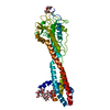 7p59C  7p5aC 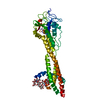 7p5dC  6elcS S: Starting model for refinement C: citing same article ( |
|---|---|
| Similar structure data |
-Links
 PDBj
PDBj

- Assembly
| Deposited unit | 
| |||||||||||||||||||||||||||||||||
|---|---|---|---|---|---|---|---|---|---|---|---|---|---|---|---|---|---|---|---|---|---|---|---|---|---|---|---|---|---|---|---|---|---|---|
| 1 |
| |||||||||||||||||||||||||||||||||
| 2 | 
| |||||||||||||||||||||||||||||||||
| Unit cell |
| |||||||||||||||||||||||||||||||||
| Components on special symmetry positions |
|
-Components
| #1: Protein | Mass: 53686.113 Da / Num. of mol.: 1 Source method: isolated from a genetically manipulated source Details: Serine319 from the original 6ELC structure has been mutated to an Alanine. Source: (gene. exp.) |
|---|---|
| #2: Polysaccharide | alpha-D-mannopyranose-(1-2)-alpha-D-mannopyranose-(1-3)-[alpha-D-mannopyranose-(1-6)]beta-D- ...alpha-D-mannopyranose-(1-2)-alpha-D-mannopyranose-(1-3)-[alpha-D-mannopyranose-(1-6)]beta-D-mannopyranose-(1-4)-2-acetamido-2-deoxy-beta-D-glucopyranose-(1-4)-2-acetamido-2-deoxy-beta-D-glucopyranose Source method: isolated from a genetically manipulated source |
| #3: Sugar | ChemComp-GLC /   Type: D-saccharide, alpha linking / Mass: 180.156 Da / Num. of mol.: 1 / Source method: obtained synthetically / Formula: C6H12O6 / Feature type: SUBJECT OF INVESTIGATION Type: D-saccharide, alpha linking / Mass: 180.156 Da / Num. of mol.: 1 / Source method: obtained synthetically / Formula: C6H12O6 / Feature type: SUBJECT OF INVESTIGATION |
| #4: Water | ChemComp-HOH /  Mass: 18.015 Da / Num. of mol.: 497 / Source method: isolated from a natural source / Formula: H2O Mass: 18.015 Da / Num. of mol.: 497 / Source method: isolated from a natural source / Formula: H2O |
| Has ligand of interest | Y |
| Has protein modification | Y |
-Experimental details
-Experiment
| Experiment | Method: X-RAY DIFFRACTION / Number of used crystals: 1 |
|---|
- Sample preparation
| Crystal grow | Temperature: 295 K / Method: vapor diffusion, hanging drop / Details: 25% PEG 3350, 300mM NaCl, 100mM HEPES, pH 7.5 |
|---|
-Data collection
| Diffraction | Mean temperature: 100 K / Serial crystal experiment: N |
|---|---|
| Diffraction source | Source: SYNCHROTRON / Site: Diamond  / Beamline: I03 / Wavelength: 0.9763 Å / Beamline: I03 / Wavelength: 0.9763 Å |
| Detector | Type: DECTRIS EIGER2 XE 16M / Detector: PIXEL / Date: Jan 26, 2020 |
| Radiation | Protocol: SINGLE WAVELENGTH / Monochromatic (M) / Laue (L): M / Scattering type: x-ray |
| Radiation wavelength | Wavelength: 0.9763 Å / Relative weight: 1 |
| Reflection | Resolution: 1.13→40.8 Å / Num. obs: 132433 / % possible obs: 96.6 % / Redundancy: 39 % / Biso Wilson estimate: 17.4 Å2 / CC1/2: 1 / CC star: 1 / Rmerge(I) obs: 0.07786 / Rpim(I) all: 0.0125 / Rrim(I) all: 0.07886 / Net I/σ(I): 23.97 |
| Reflection shell | Resolution: 1.13→1.17 Å / Redundancy: 28.4 % / Rmerge(I) obs: 6.968 / Mean I/σ(I) obs: 0.42 / Num. unique obs: 8709 / CC1/2: 0.169 / CC star: 0.538 / Rpim(I) all: 1.324 / Rrim(I) all: 7.094 / % possible all: 66.13 |
- Processing
| Software |
| |||||||||||||||||||||||||||||||||||||||||||||||||||||||||||||||||||||||||||||||||||||||||||||||||||||||||||||||||||||||||||||||||||||||||||||||||||||||||||||||||||||||||||||||||||||||||||||||||||||||||||||||||||||||||
|---|---|---|---|---|---|---|---|---|---|---|---|---|---|---|---|---|---|---|---|---|---|---|---|---|---|---|---|---|---|---|---|---|---|---|---|---|---|---|---|---|---|---|---|---|---|---|---|---|---|---|---|---|---|---|---|---|---|---|---|---|---|---|---|---|---|---|---|---|---|---|---|---|---|---|---|---|---|---|---|---|---|---|---|---|---|---|---|---|---|---|---|---|---|---|---|---|---|---|---|---|---|---|---|---|---|---|---|---|---|---|---|---|---|---|---|---|---|---|---|---|---|---|---|---|---|---|---|---|---|---|---|---|---|---|---|---|---|---|---|---|---|---|---|---|---|---|---|---|---|---|---|---|---|---|---|---|---|---|---|---|---|---|---|---|---|---|---|---|---|---|---|---|---|---|---|---|---|---|---|---|---|---|---|---|---|---|---|---|---|---|---|---|---|---|---|---|---|---|---|---|---|---|---|---|---|---|---|---|---|---|---|---|---|---|---|---|---|---|
| Refinement | Method to determine structure: MOLECULAR REPLACEMENT Starting model: 6ELC Resolution: 1.13→40.8 Å / SU ML: 0.16 / Cross valid method: FREE R-VALUE / σ(F): 1.33 / Phase error: 24.84 / Stereochemistry target values: ML
| |||||||||||||||||||||||||||||||||||||||||||||||||||||||||||||||||||||||||||||||||||||||||||||||||||||||||||||||||||||||||||||||||||||||||||||||||||||||||||||||||||||||||||||||||||||||||||||||||||||||||||||||||||||||||
| Solvent computation | Shrinkage radii: 0.9 Å / VDW probe radii: 1.11 Å / Solvent model: FLAT BULK SOLVENT MODEL | |||||||||||||||||||||||||||||||||||||||||||||||||||||||||||||||||||||||||||||||||||||||||||||||||||||||||||||||||||||||||||||||||||||||||||||||||||||||||||||||||||||||||||||||||||||||||||||||||||||||||||||||||||||||||
| Displacement parameters | Biso max: 79.41 Å2 / Biso mean: 25.6297 Å2 / Biso min: 12.93 Å2 | |||||||||||||||||||||||||||||||||||||||||||||||||||||||||||||||||||||||||||||||||||||||||||||||||||||||||||||||||||||||||||||||||||||||||||||||||||||||||||||||||||||||||||||||||||||||||||||||||||||||||||||||||||||||||
| Refinement step | Cycle: final / Resolution: 1.13→40.8 Å
| |||||||||||||||||||||||||||||||||||||||||||||||||||||||||||||||||||||||||||||||||||||||||||||||||||||||||||||||||||||||||||||||||||||||||||||||||||||||||||||||||||||||||||||||||||||||||||||||||||||||||||||||||||||||||
| LS refinement shell | Refine-ID: X-RAY DIFFRACTION / Rfactor Rfree error: 0 / Total num. of bins used: 30
| |||||||||||||||||||||||||||||||||||||||||||||||||||||||||||||||||||||||||||||||||||||||||||||||||||||||||||||||||||||||||||||||||||||||||||||||||||||||||||||||||||||||||||||||||||||||||||||||||||||||||||||||||||||||||
| Refinement TLS params. | Method: refined / Refine-ID: X-RAY DIFFRACTION
| |||||||||||||||||||||||||||||||||||||||||||||||||||||||||||||||||||||||||||||||||||||||||||||||||||||||||||||||||||||||||||||||||||||||||||||||||||||||||||||||||||||||||||||||||||||||||||||||||||||||||||||||||||||||||
| Refinement TLS group |
|
