 Movie
Movie Controller

[English] 日本語
 Yorodumi
- PDB-7p4v: GlnK1 from Methanothermococcus thermolithotrophicus with dADP at ... -
+ Open data

- Basic information
| Entry | Database: PDB / ID: 7p4v | ||||||
|---|---|---|---|---|---|---|---|
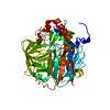
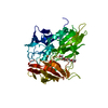
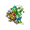
| Title | GlnK1 from Methanothermococcus thermolithotrophicus with dADP at a resolution of 1.94 A | ||||||
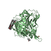
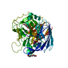
 Components Components | GlnK1 from Methanothermococcus thermolithotrophicus | ||||||
 Keywords Keywords | SIGNALING PROTEIN / PII-family / Methanococcales / methanogenic archaea / hydrogenotrophs / protein regulation / inhibitor / T-loop / conformational change / dADP / 2-oxoglutarate / nitrogen metabolism | ||||||
| Function / homology | 2'-DEOXYADENOSINE-5'-DIPHOSPHATE Function and homology information Function and homology information | ||||||
| Biological species |  Methanothermococcus thermolithotrophicus DSM 2095 (archaea) Methanothermococcus thermolithotrophicus DSM 2095 (archaea) | ||||||
| Method |  X-RAY DIFFRACTION / SYNCHROTRON / MOLECULAR REPLACEMENT / Resolution: 1.94 Å X-RAY DIFFRACTION / SYNCHROTRON / MOLECULAR REPLACEMENT / Resolution: 1.94 Å | ||||||
 Authors Authors | Mueller, M.-C. / Wagner, T. | ||||||
| Funding support |  Germany, 1items Germany, 1items
| ||||||
 Citation Citation | Journal: Int J Mol Sci / Year: 2021 Title: The Oxoglutarate Binding Site and Regulatory Mechanism Are Conserved in Ammonium Transporter Inhibitors GlnKs from Methanococcales . Authors: Muller, M.C. / Wagner, T. | ||||||
| History |
|
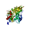
- Structure visualization
| Structure viewer | Molecule: MolmilJmol/JSmol |
|---|
- Downloads & links
-Download
| PDBx/mmCIF format | 7p4v.cif.gz | 62.4 KB | Display | PDBx/mmCIF format |
|---|---|---|---|---|
| PDB format | pdb7p4v.ent.gz | 44.5 KB | Display | PDB format |
| PDBx/mmJSON format | 7p4v.json.gz | Tree view | PDBx/mmJSON format | |
| Others |  Other downloads Other downloads |
-Validation report
| Arichive directory | https://data.pdbj.org/pub/pdb/validation_reports/p4/7p4vftp://data.pdbj.org/pub/pdb/validation_reports/p4/7p4v | HTTPS FTP |
|---|
-Related structure data
| Related structure data | 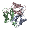 7p4yC  7p50C 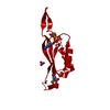 7p52C 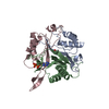 2j9dS S: Starting model for refinement C: citing same article ( |
|---|---|
| Similar structure data |


-Links
 PDBj
PDBj- Assembly
| Deposited unit | 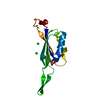
| ||||||||||||
|---|---|---|---|---|---|---|---|---|---|---|---|---|---|
| 1 | 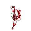
| ||||||||||||
| Unit cell |
| ||||||||||||
| Components on special symmetry positions |
|
-Components
| #1: Protein | Mass: 14648.796 Da / Num. of mol.: 1 Source method: isolated from a genetically manipulated source Details: / Source: (gene. exp.) Methanothermococcus thermolithotrophicus DSM 2095 (archaea)Strain: DSM 2095 / Tissue: / / Cell: / / Cell line: / / Gene: glnk1 / Organ: / / Variant: / / Plasmid: pET-28a(+) / Cell (production host): / / Cell line (production host): / / Organ (production host): / / Production host:  | ||||
|---|---|---|---|---|---|
| #2: Chemical | ChemComp-DAT /   Mass: 411.202 Da / Num. of mol.: 1 / Source method: obtained synthetically / Formula: C10H15N5O9P2 / Feature type: SUBJECT OF INVESTIGATION Mass: 411.202 Da / Num. of mol.: 1 / Source method: obtained synthetically / Formula: C10H15N5O9P2 / Feature type: SUBJECT OF INVESTIGATION | ||||
| #3: Chemical |   Mass: 35.453 Da / Num. of mol.: 2 / Source method: obtained synthetically / Formula: Cl Mass: 35.453 Da / Num. of mol.: 2 / Source method: obtained synthetically / Formula: Cl#4: Water | ChemComp-HOH / |  Mass: 18.015 Da / Num. of mol.: 23 / Source method: isolated from a natural source / Formula: H2O Mass: 18.015 Da / Num. of mol.: 23 / Source method: isolated from a natural source / Formula: H2OHas ligand of interest | Y | |
-Experimental details
-Experiment
| Experiment | Method: X-RAY DIFFRACTION / Number of used crystals: 1 |
|---|
- Sample preparation
| Crystal | Density Matthews: 3.48 Å3/Da / Density % sol: 64.69 % / Description: transparent hexagonal rod |
|---|---|
| Crystal grow | Temperature: 291.15 K / Method: vapor diffusion, sitting drop / pH: 6.5 Details: GlnK1 crystallized at a concentration of 33 mg/ml in 25 mM TrisHCl pH 7.6, 10% glycerol, 2mM dithiothreitol and 500mM NaCl . Drop of 0.6 ul of protein was mixed with 0.6 ul of the ...Details: GlnK1 crystallized at a concentration of 33 mg/ml in 25 mM TrisHCl pH 7.6, 10% glycerol, 2mM dithiothreitol and 500mM NaCl . Drop of 0.6 ul of protein was mixed with 0.6 ul of the crystallization solution in a 96-Well MRC 2-Drop Crystallization Plates in polystyrene (SWISSCI). The reservoir contained 90 ul of the following crystallization solution: 35 % w/v Pentaerythritol propoxylate (17/8 PO/OH), 100 mM MES pH 6.5, 200 mM Ammonium sulfate. |
-Data collection
| Diffraction | Mean temperature: 100 K / Serial crystal experiment: N |
|---|---|
| Diffraction source | Source: SYNCHROTRON / Site: SLS  / Beamline: X06DA / Wavelength: 1.00003 Å / Beamline: X06DA / Wavelength: 1.00003 Å |
| Detector | Type: DECTRIS PILATUS 2M-F / Detector: PIXEL / Date: Dec 17, 2019 |
| Radiation | Protocol: SINGLE WAVELENGTH / Monochromatic (M) / Laue (L): M / Scattering type: x-ray |
| Radiation wavelength | Wavelength: 1.00003 Å / Relative weight: 1 |
| Reflection | Resolution: 1.94→75.601 Å / Num. obs: 10108 / % possible obs: 89.1 % / Redundancy: 19.3 % / CC1/2: 0.999 / Rmerge(I) obs: 0.053 / Rpim(I) all: 0.013 / Rrim(I) all: 0.055 / Net I/σ(I): 27.3 |
| Reflection shell | Resolution: 1.94→2.148 Å / Redundancy: 18.6 % / Rmerge(I) obs: 1.711 / Mean I/σ(I) obs: 1.8 / Num. unique obs: 502 / CC1/2: 0.773 / Rpim(I) all: 0.403 / Rrim(I) all: 1.759 / % possible all: 60.2 |
- Processing
| Software |
| |||||||||||||||||||||||||||||||||||||||||||||||||||||||||||||||||||||||||||||||||||||||||||||||||||||||||||||||||||||||||||||||||||||||||||||||||||||||||||||||||||||||||||||||
|---|---|---|---|---|---|---|---|---|---|---|---|---|---|---|---|---|---|---|---|---|---|---|---|---|---|---|---|---|---|---|---|---|---|---|---|---|---|---|---|---|---|---|---|---|---|---|---|---|---|---|---|---|---|---|---|---|---|---|---|---|---|---|---|---|---|---|---|---|---|---|---|---|---|---|---|---|---|---|---|---|---|---|---|---|---|---|---|---|---|---|---|---|---|---|---|---|---|---|---|---|---|---|---|---|---|---|---|---|---|---|---|---|---|---|---|---|---|---|---|---|---|---|---|---|---|---|---|---|---|---|---|---|---|---|---|---|---|---|---|---|---|---|---|---|---|---|---|---|---|---|---|---|---|---|---|---|---|---|---|---|---|---|---|---|---|---|---|---|---|---|---|---|---|---|---|---|
| Refinement | Method to determine structure: MOLECULAR REPLACEMENT Starting model: 2J9D Resolution: 1.94→39.3 Å / SU ML: 0.19 / Cross valid method: THROUGHOUT / σ(F): 1.34 / Phase error: 36.79 / Stereochemistry target values: ML Details: The last refinement cycle was performed with hydrogens in riding position
| |||||||||||||||||||||||||||||||||||||||||||||||||||||||||||||||||||||||||||||||||||||||||||||||||||||||||||||||||||||||||||||||||||||||||||||||||||||||||||||||||||||||||||||||
| Solvent computation | Shrinkage radii: 0.9 Å / VDW probe radii: 1.11 Å / Solvent model: FLAT BULK SOLVENT MODEL | |||||||||||||||||||||||||||||||||||||||||||||||||||||||||||||||||||||||||||||||||||||||||||||||||||||||||||||||||||||||||||||||||||||||||||||||||||||||||||||||||||||||||||||||
| Displacement parameters | Biso max: 130.32 Å2 / Biso mean: 62.7293 Å2 / Biso min: 32.23 Å2 | |||||||||||||||||||||||||||||||||||||||||||||||||||||||||||||||||||||||||||||||||||||||||||||||||||||||||||||||||||||||||||||||||||||||||||||||||||||||||||||||||||||||||||||||
| Refinement step | Cycle: final / Resolution: 1.94→39.3 Å
| |||||||||||||||||||||||||||||||||||||||||||||||||||||||||||||||||||||||||||||||||||||||||||||||||||||||||||||||||||||||||||||||||||||||||||||||||||||||||||||||||||||||||||||||
| LS refinement shell | Refine-ID: X-RAY DIFFRACTION / Rfactor Rfree error: 0 / Total num. of bins used: 4
| |||||||||||||||||||||||||||||||||||||||||||||||||||||||||||||||||||||||||||||||||||||||||||||||||||||||||||||||||||||||||||||||||||||||||||||||||||||||||||||||||||||||||||||||
| Refinement TLS params. | Method: refined / Refine-ID: X-RAY DIFFRACTION
| |||||||||||||||||||||||||||||||||||||||||||||||||||||||||||||||||||||||||||||||||||||||||||||||||||||||||||||||||||||||||||||||||||||||||||||||||||||||||||||||||||||||||||||||
| Refinement TLS group |
|
