 Movie
Movie Controller

[English] 日本語
 Yorodumi
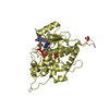
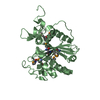
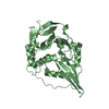
- PDB-2j9d: Structure of GlnK1 with bound effectors indicates regulatory mech... -
+ Open data

- Basic information
| Entry | Database: PDB / ID: 2j9d | ||||||
|---|---|---|---|---|---|---|---|
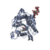
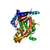
| Title | Structure of GlnK1 with bound effectors indicates regulatory mechanism for ammonia uptake | ||||||
 Components Components | (HYPOTHETICAL NITROGEN REGULATORY PII-LIKE PROTEIN ...) x 2 | ||||||
 Keywords Keywords | MEMBRANE TRANSPORT / EM SINGLE PARTICLE / NITROGEN METABOLISM / SIGNALLING / TRANSCRIPTION / HYPOTHETICAL PROTEIN / TRANSCRIPTION REGULATION | ||||||
| Function / homology |  Function and homology information Function and homology informationregulation of nitrogen utilization / enzyme regulator activity / ATP binding / cytosol Similarity search - Function | ||||||
| Biological species |   METHANOCOCCUS JANNASCHII (archaea) METHANOCOCCUS JANNASCHII (archaea) | ||||||
| Method |  X-RAY DIFFRACTION / SYNCHROTRON / MOLECULAR REPLACEMENT / Resolution: 2.1 Å X-RAY DIFFRACTION / SYNCHROTRON / MOLECULAR REPLACEMENT / Resolution: 2.1 Å | ||||||
 Authors Authors | Yildiz, O. / Kalthoff, C. / Raunser, S. / Kuehlbrandt, W. | ||||||
 Citation Citation | Journal: Embo J. / Year: 2007 Title: Structure of Glnk1 with Bound Effectors Indicates Regulatory Mechanism for Ammonia Uptake. Authors: Yildiz, O. / Kalthoff, C. / Raunser, S. / Kuhlbrandt, W. | ||||||
| History |
| ||||||
| Remark 700 | SHEET THE SHEET STRUCTURE OF THIS MOLECULE IS BIFURCATED. IN ORDER TO REPRESENT THIS FEATURE IN ... SHEET THE SHEET STRUCTURE OF THIS MOLECULE IS BIFURCATED. IN ORDER TO REPRESENT THIS FEATURE IN THE SHEET RECORDS BELOW, TWO SHEETS ARE DEFINED. |
- Structure visualization
| Structure viewer | Molecule: MolmilJmol/JSmol |
|---|
- Downloads & links
-Download
| PDBx/mmCIF format | 2j9d.cif.gz | 274.3 KB | Display | PDBx/mmCIF format |
|---|---|---|---|---|
| PDB format | pdb2j9d.ent.gz | 223.2 KB | Display | PDB format |
| PDBx/mmJSON format | 2j9d.json.gz | Tree view | PDBx/mmJSON format | |
| Others |  Other downloads Other downloads |
-Validation report
| Arichive directory | https://data.pdbj.org/pub/pdb/validation_reports/j9/2j9dftp://data.pdbj.org/pub/pdb/validation_reports/j9/2j9d | HTTPS FTP |
|---|
-Related structure data
| Related structure data | 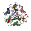 2j9cSC 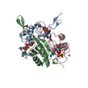 2j9eC S: Starting model for refinement C: citing same article ( |
|---|---|
| Similar structure data |


-Links
 PDBj
PDBj


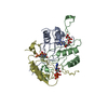
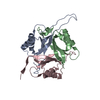
- Assembly
| Deposited unit | 
| ||||||||
|---|---|---|---|---|---|---|---|---|---|
| 1 | 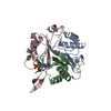
| ||||||||
| 2 | 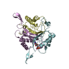
| ||||||||
| 3 | 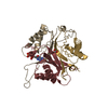
| ||||||||
| 4 | 
| ||||||||
| Unit cell |
|
-Components
-HYPOTHETICAL NITROGEN REGULATORY PII-LIKE PROTEIN ... , 2 types, 12 molecules ABCDFGHIJKLE
| #1: Protein | Mass: 13311.469 Da / Num. of mol.: 11 Source method: isolated from a genetically manipulated source Source: (gene. exp.) METHANOCOCCUS JANNASCHII (archaea) / Strain: AMJFT37 / Plasmid: PET28-D2 / Production host:  #2: Protein | | Mass: 13327.425 Da / Num. of mol.: 1 Source method: isolated from a genetically manipulated source Source: (gene. exp.) METHANOCOCCUS JANNASCHII (archaea) / Strain: AMJFT37 / Plasmid: PET28-D2 / Production host: |
|---|
-Non-polymers , 5 types, 705 molecules 


| #3: Chemical | ChemComp-ACT /  Mass: 59.044 Da / Num. of mol.: 5 / Source method: obtained synthetically / Formula: C2H3O2 Mass: 59.044 Da / Num. of mol.: 5 / Source method: obtained synthetically / Formula: C2H3O2#4: Chemical | ChemComp-ADP /  Mass: 427.201 Da / Num. of mol.: 4 / Source method: obtained synthetically / Formula: C10H15N5O10P2 / Comment: ADP, energy-carrying molecule*YM Mass: 427.201 Da / Num. of mol.: 4 / Source method: obtained synthetically / Formula: C10H15N5O10P2 / Comment: ADP, energy-carrying molecule*YM#5: Chemical | ChemComp-AMP / |  Mass: 347.221 Da / Num. of mol.: 1 / Source method: obtained synthetically / Formula: C10H14N5O7P / Comment: AMP*YM Mass: 347.221 Da / Num. of mol.: 1 / Source method: obtained synthetically / Formula: C10H14N5O7P / Comment: AMP*YM#6: Chemical | ChemComp-CL / |  Mass: 35.453 Da / Num. of mol.: 1 / Source method: obtained synthetically / Formula: Cl Mass: 35.453 Da / Num. of mol.: 1 / Source method: obtained synthetically / Formula: Cl#7: Water | ChemComp-HOH / | Mass: 18.015 Da / Num. of mol.: 694 / Source method: isolated from a natural source / Formula: H2O |
|---|
-Experimental details
-Experiment
| Experiment | Method: X-RAY DIFFRACTION / Number of used crystals: 1 |
|---|
- Sample preparation
| Crystal | Density Matthews: 2.17 Å3/Da / Density % sol: 43.41 % |
|---|---|
| Crystal grow | pH: 7.5 / Details: pH 7.50 |
-Data collection
| Diffraction | Mean temperature: 100 K |
|---|---|
| Diffraction source | Source: SYNCHROTRON / Site: ESRF  / Beamline: ID14-1 / Wavelength: 0.934 / Beamline: ID14-1 / Wavelength: 0.934 |
| Detector | Type: ADSC CCD / Detector: CCD / Date: May 9, 2004 |
| Radiation | Protocol: SINGLE WAVELENGTH / Monochromatic (M) / Laue (L): M / Scattering type: x-ray |
| Radiation wavelength | Wavelength: 0.934 Å / Relative weight: 1 |
| Reflection | Resolution: 2.1→20 Å / Num. obs: 80980 / % possible obs: 99 % / Observed criterion σ(I): 3 / Redundancy: 4.6 % / Rmerge(I) obs: 0.16 / Net I/σ(I): 11.3 |
| Reflection shell | Resolution: 2.1→2.2 Å / Redundancy: 4.6 % / Rmerge(I) obs: 0.53 / Mean I/σ(I) obs: 3.06 / % possible all: 98.8 |
- Processing
| Software |
| ||||||||||||||||||||||||||||||||||||||||||||||||||||||||||||||||||||||||||||||||||||||||||||||||||||||||||||||||||||||||||||||||||||||||||||||||||||||||||||||||||||||||||||||||||||||
|---|---|---|---|---|---|---|---|---|---|---|---|---|---|---|---|---|---|---|---|---|---|---|---|---|---|---|---|---|---|---|---|---|---|---|---|---|---|---|---|---|---|---|---|---|---|---|---|---|---|---|---|---|---|---|---|---|---|---|---|---|---|---|---|---|---|---|---|---|---|---|---|---|---|---|---|---|---|---|---|---|---|---|---|---|---|---|---|---|---|---|---|---|---|---|---|---|---|---|---|---|---|---|---|---|---|---|---|---|---|---|---|---|---|---|---|---|---|---|---|---|---|---|---|---|---|---|---|---|---|---|---|---|---|---|---|---|---|---|---|---|---|---|---|---|---|---|---|---|---|---|---|---|---|---|---|---|---|---|---|---|---|---|---|---|---|---|---|---|---|---|---|---|---|---|---|---|---|---|---|---|---|---|---|
| Refinement | Method to determine structure: MOLECULAR REPLACEMENT Starting model: PDB ENTRY 2J9C Resolution: 2.1→19.78 Å / Cor.coef. Fo:Fc: 0.937 / Cor.coef. Fo:Fc free: 0.896 / SU B: 5.657 / SU ML: 0.154 / Cross valid method: THROUGHOUT / ESU R: 0.251 / ESU R Free: 0.213 / Stereochemistry target values: MAXIMUM LIKELIHOOD / Details: HYDROGENS HAVE BEEN ADDED IN THE RIDING POSITIONS.
| ||||||||||||||||||||||||||||||||||||||||||||||||||||||||||||||||||||||||||||||||||||||||||||||||||||||||||||||||||||||||||||||||||||||||||||||||||||||||||||||||||||||||||||||||||||||
| Solvent computation | Ion probe radii: 0.8 Å / Shrinkage radii: 0.8 Å / VDW probe radii: 1.4 Å / Solvent model: BABINET MODEL WITH MASK | ||||||||||||||||||||||||||||||||||||||||||||||||||||||||||||||||||||||||||||||||||||||||||||||||||||||||||||||||||||||||||||||||||||||||||||||||||||||||||||||||||||||||||||||||||||||
| Displacement parameters | Biso mean: 30.31 Å2
| ||||||||||||||||||||||||||||||||||||||||||||||||||||||||||||||||||||||||||||||||||||||||||||||||||||||||||||||||||||||||||||||||||||||||||||||||||||||||||||||||||||||||||||||||||||||
| Refinement step | Cycle: LAST / Resolution: 2.1→19.78 Å
| ||||||||||||||||||||||||||||||||||||||||||||||||||||||||||||||||||||||||||||||||||||||||||||||||||||||||||||||||||||||||||||||||||||||||||||||||||||||||||||||||||||||||||||||||||||||
| Refine LS restraints |
|