 Movie
Movie Controller

[English] 日本語
 Yorodumi
- PDB-7p4f: Crystal Structure of Monoamine Oxidase B in complex with inhibitor 1 -
+ Open data

- Basic information
| Entry | Database: PDB / ID: 7p4f | ||||||
|---|---|---|---|---|---|---|---|
| Title | Crystal Structure of Monoamine Oxidase B in complex with inhibitor 1 | ||||||
 Components Components | Amine oxidase [flavin-containing] B | ||||||
 Keywords Keywords | FLAVOPROTEIN / monoamine oxidase / drug target / neurodegeneration / mitochondrial membrane | ||||||
| Function / homology |  Function and homology information Function and homology informationBiogenic amines are oxidatively deaminated to aldehydes by MAOA and MAOB / monoamine oxidase / monoamine oxidase activity / primary-amine oxidase / primary methylamine oxidase activity / dopamine catabolic process / mitochondrial envelope / hydrogen peroxide biosynthetic process / substantia nigra development / flavin adenine dinucleotide binding ...Biogenic amines are oxidatively deaminated to aldehydes by MAOA and MAOB / monoamine oxidase / monoamine oxidase activity / primary-amine oxidase / primary methylamine oxidase activity / dopamine catabolic process / mitochondrial envelope / hydrogen peroxide biosynthetic process / substantia nigra development / flavin adenine dinucleotide binding / mitochondrial outer membrane / electron transfer activity / mitochondrion Similarity search - Function | ||||||
| Biological species |  Homo sapiens (human) Homo sapiens (human) | ||||||
| Method |  X-RAY DIFFRACTION / SYNCHROTRON / MOLECULAR REPLACEMENT / Resolution: 2.3 Å X-RAY DIFFRACTION / SYNCHROTRON / MOLECULAR REPLACEMENT / Resolution: 2.3 Å | ||||||
 Authors Authors | Iacovino, L.G. / Binda, C. / Pisani, L. | ||||||
 Citation Citation | Journal: Acs Med.Chem.Lett. / Year: 2022 Title: Dual Reversible Coumarin Inhibitors Mutually Bound to Monoamine Oxidase B and Acetylcholinesterase Crystal Structures. Authors: Ekstrom, F. / Gottinger, A. / Forsgren, N. / Catto, M. / Iacovino, L.G. / Pisani, L. / Binda, C. | ||||||
| History |
|
- Structure visualization
| Structure viewer | Molecule: MolmilJmol/JSmol |
|---|
- Downloads & links
-Download
| PDBx/mmCIF format | 7p4f.cif.gz | 221 KB | Display | PDBx/mmCIF format |
|---|---|---|---|---|
| PDB format | pdb7p4f.ent.gz | 175.5 KB | Display | PDB format |
| PDBx/mmJSON format | 7p4f.json.gz | Tree view | PDBx/mmJSON format | |
| Others |  Other downloads Other downloads |
-Validation report
| Arichive directory | https://data.pdbj.org/pub/pdb/validation_reports/p4/7p4fftp://data.pdbj.org/pub/pdb/validation_reports/p4/7p4f | HTTPS FTP |
|---|
-Related structure data
| Related structure data |  7p4hC 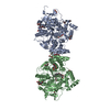 7qakC  7qb4C 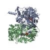 2v5zS S: Starting model for refinement C: citing same article ( |
|---|---|
| Similar structure data |
-Links
 PDBj
PDBj

- Assembly
| Deposited unit | 
| |||||||||
|---|---|---|---|---|---|---|---|---|---|---|
| 1 |
| |||||||||
| Unit cell |
| |||||||||
| Components on special symmetry positions |
|
-Components
| #1: Protein | Mass: 58837.730 Da / Num. of mol.: 2 Source method: isolated from a genetically manipulated source Source: (gene. exp.) Homo sapiens (human) / Gene: MAOB / Production host:  Komagataella pastoris (fungus) / References: UniProt: P27338, monoamine oxidase Komagataella pastoris (fungus) / References: UniProt: P27338, monoamine oxidase#2: Chemical |   Mass: 785.550 Da / Num. of mol.: 2 / Source method: obtained synthetically / Formula: C27H33N9O15P2 / Comment: FAD*YM Mass: 785.550 Da / Num. of mol.: 2 / Source method: obtained synthetically / Formula: C27H33N9O15P2 / Comment: FAD*YM#3: Chemical |   Mass: 415.481 Da / Num. of mol.: 2 / Source method: obtained synthetically / Formula: C26H25NO4 Mass: 415.481 Da / Num. of mol.: 2 / Source method: obtained synthetically / Formula: C26H25NO4#4: Chemical |   Mass: 336.554 Da / Num. of mol.: 2 / Source method: obtained synthetically / Formula: C17H38NO3S Mass: 336.554 Da / Num. of mol.: 2 / Source method: obtained synthetically / Formula: C17H38NO3S#5: Water | ChemComp-HOH / |  Mass: 18.015 Da / Num. of mol.: 383 / Source method: isolated from a natural source / Formula: H2O Mass: 18.015 Da / Num. of mol.: 383 / Source method: isolated from a natural source / Formula: H2OHas protein modification | Y | |
|---|
-Experimental details
-Experiment
| Experiment | Method: X-RAY DIFFRACTION / Number of used crystals: 1 |
|---|
- Sample preparation
| Crystal | Density Matthews: 2.67 Å3/Da / Density % sol: 53.99 % |
|---|---|
| Crystal grow | Temperature: 277 K / Method: vapor diffusion / Details: PEG 4000, lithium sulfate, ADA buffer |
-Data collection
| Diffraction | Mean temperature: 100 K / Serial crystal experiment: N |
|---|---|
| Diffraction source | Source: SYNCHROTRON / Site: SLS  / Beamline: X06DA / Wavelength: 0.9786 Å / Beamline: X06DA / Wavelength: 0.9786 Å |
| Detector | Type: DECTRIS PILATUS 2M / Detector: PIXEL / Date: May 14, 2020 |
| Radiation | Protocol: SINGLE WAVELENGTH / Monochromatic (M) / Laue (L): M / Scattering type: x-ray |
| Radiation wavelength | Wavelength: 0.9786 Å / Relative weight: 1 |
| Reflection | Resolution: 2.3→47.31 Å / Num. obs: 54746 / % possible obs: 99.6 % / Redundancy: 5.3 % / CC1/2: 0.99 / Net I/σ(I): 9.3 |
| Reflection shell | Resolution: 2.3→2.36 Å / Num. unique obs: 4014 / CC1/2: 0.76 |
- Processing
| Software |
| ||||||||||||||||||||||||||||||||||||||||||||||||||||||||||||
|---|---|---|---|---|---|---|---|---|---|---|---|---|---|---|---|---|---|---|---|---|---|---|---|---|---|---|---|---|---|---|---|---|---|---|---|---|---|---|---|---|---|---|---|---|---|---|---|---|---|---|---|---|---|---|---|---|---|---|---|---|---|
| Refinement | Method to determine structure: MOLECULAR REPLACEMENT Starting model: 2v5z Resolution: 2.3→47.31 Å / Cor.coef. Fo:Fc: 0.956 / Cor.coef. Fo:Fc free: 0.918 / SU B: 5.734 / SU ML: 0.138 / Cross valid method: THROUGHOUT / σ(F): 0 / ESU R: 0.24 / ESU R Free: 0.195 / Stereochemistry target values: MAXIMUM LIKELIHOOD Details: HYDROGENS HAVE BEEN ADDED IN THE RIDING POSITIONS U VALUES : REFINED INDIVIDUALLY
| ||||||||||||||||||||||||||||||||||||||||||||||||||||||||||||
| Solvent computation | Ion probe radii: 0.8 Å / Shrinkage radii: 0.8 Å / VDW probe radii: 1.2 Å / Solvent model: MASK | ||||||||||||||||||||||||||||||||||||||||||||||||||||||||||||
| Displacement parameters | Biso max: 99.17 Å2 / Biso mean: 28.935 Å2 / Biso min: 10.84 Å2
| ||||||||||||||||||||||||||||||||||||||||||||||||||||||||||||
| Refinement step | Cycle: final / Resolution: 2.3→47.31 Å
| ||||||||||||||||||||||||||||||||||||||||||||||||||||||||||||
| Refine LS restraints |
| ||||||||||||||||||||||||||||||||||||||||||||||||||||||||||||
| LS refinement shell | Resolution: 2.3→2.36 Å / Rfactor Rfree error: 0 / Total num. of bins used: 20
|
