Movie
Movie Controller


+ Open data

- Basic information
| Entry | Database: PDB / ID: 7p30 | |||||||||||||||
|---|---|---|---|---|---|---|---|---|---|---|---|---|---|---|---|---|
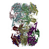
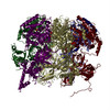
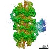
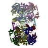
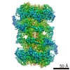
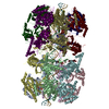
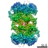
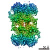
| Title | 3.0 A resolution structure of a DNA-loaded MCM double hexamer | |||||||||||||||
 Components Components |
| |||||||||||||||
 Keywords Keywords | REPLICATION / Mcm2-7 helicase / nucleoprotein complex / AAA+ ATPase / DNA replication | |||||||||||||||
| Function / homology |  Function and homology information Function and homology informationMCM core complex / Assembly of the pre-replicative complex / Switching of origins to a post-replicative state / MCM complex binding / nuclear DNA replication / premeiotic DNA replication / pre-replicative complex assembly involved in nuclear cell cycle DNA replication / Activation of the pre-replicative complex / mitotic DNA replication / nuclear pre-replicative complex ...MCM core complex / Assembly of the pre-replicative complex / Switching of origins to a post-replicative state / MCM complex binding / nuclear DNA replication / premeiotic DNA replication / pre-replicative complex assembly involved in nuclear cell cycle DNA replication / Activation of the pre-replicative complex / mitotic DNA replication / nuclear pre-replicative complex / CMG complex / DNA replication preinitiation complex / Activation of ATR in response to replication stress / double-strand break repair via break-induced replication / MCM complex / mitotic DNA replication initiation / silent mating-type cassette heterochromatin formation / single-stranded DNA helicase activity / regulation of DNA-templated DNA replication initiation / DNA strand elongation involved in DNA replication / nuclear replication fork / DNA replication origin binding / DNA replication initiation / subtelomeric heterochromatin formation / DNA helicase activity / helicase activity / transcription elongation by RNA polymerase II / peroxisome / heterochromatin formation / single-stranded DNA binding / DNA helicase / DNA replication / chromosome, telomeric region / chromatin binding / DNA damage response / ATP hydrolysis activity / zinc ion binding / nucleoplasm / ATP binding / nucleus / cytoplasm Similarity search - Function | |||||||||||||||
| Biological species |  | |||||||||||||||
| Method | ELECTRON MICROSCOPY / single particle reconstruction / cryo EM / Resolution: 3 Å | |||||||||||||||
 Authors Authors | Greiwe, J.F. / Miller, T.C.R. / Martino, F. / Costa, A. | |||||||||||||||
| Funding support | European Union, 4items
| |||||||||||||||
 Citation Citation | Journal: Nat Struct Mol Biol / Year: 2022 Title: Structural mechanism for the selective phosphorylation of DNA-loaded MCM double hexamers by the Dbf4-dependent kinase. Authors: Julia F Greiwe / Thomas C R Miller / Julia Locke / Fabrizio Martino / Steven Howell / Anne Schreiber / Andrea Nans / John F X Diffley / Alessandro Costa /    Abstract: Loading of the eukaryotic replicative helicase onto replication origins involves two MCM hexamers forming a double hexamer (DH) around duplex DNA. During S phase, helicase activation requires MCM ...Loading of the eukaryotic replicative helicase onto replication origins involves two MCM hexamers forming a double hexamer (DH) around duplex DNA. During S phase, helicase activation requires MCM phosphorylation by Dbf4-dependent kinase (DDK), comprising Cdc7 and Dbf4. DDK selectively phosphorylates loaded DHs, but how such fidelity is achieved is unknown. Here, we determine the cryogenic electron microscopy structure of Saccharomyces cerevisiae DDK in the act of phosphorylating a DH. DDK docks onto one MCM ring and phosphorylates the opposed ring. Truncation of the Dbf4 docking domain abrogates DH phosphorylation, yet Cdc7 kinase activity is unaffected. Late origin firing is blocked in response to DNA damage via Dbf4 phosphorylation by the Rad53 checkpoint kinase. DDK phosphorylation by Rad53 impairs DH phosphorylation by blockage of DDK binding to DHs, and also interferes with the Cdc7 active site. Our results explain the structural basis and regulation of the selective phosphorylation of DNA-loaded MCM DHs, which supports bidirectional replication. | |||||||||||||||
| History |
|
- Structure visualization
| Movie |
Movie viewer |
|---|---|
| Structure viewer | Molecule: MolmilJmol/JSmol |
- Downloads & links
-Download
| PDBx/mmCIF format | 7p30.cif.gz | 2.6 MB | Display | PDBx/mmCIF format |
|---|---|---|---|---|
| PDB format | pdb7p30.ent.gz | Display | PDB format | |
| PDBx/mmJSON format | 7p30.json.gz | Tree view | PDBx/mmJSON format | |
| Others |  Other downloads Other downloads |
-Validation report
| Arichive directory | https://data.pdbj.org/pub/pdb/validation_reports/p3/7p30ftp://data.pdbj.org/pub/pdb/validation_reports/p3/7p30 | HTTPS FTP |
|---|
-Related structure data
| Related structure data | 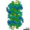 13176MC 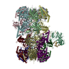 7p5zC M: map data used to model this data C: citing same article ( |
|---|---|
| Similar structure data |

-Links
 PDBj
PDBj


- Assembly
| Deposited unit | 
|
|---|---|
| 1 |
|
-Components
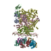
-DNA replication licensing factor ... , 5 types, 10 molecules 2A3B4C6E7F
| #1: Protein | Mass: 98911.539 Da / Num. of mol.: 2 Source method: isolated from a genetically manipulated source Source: (gene. exp.) Strain: ATCC 204508 / S288c / Gene: MCM2, YBL023C, YBL0438 / Production host: #2: Protein | Mass: 111720.242 Da / Num. of mol.: 2 Source method: isolated from a genetically manipulated source Source: (gene. exp.) Strain: ATCC 204508 / S288c / Gene: MCM3, YEL032W, SYGP-ORF23 / Production host: #3: Protein | Mass: 105138.375 Da / Num. of mol.: 2 Source method: isolated from a genetically manipulated source Source: (gene. exp.) Strain: ATCC 204508 / S288c / Gene: MCM4, CDC54, HCD21, YPR019W, YP9531.13 / Production host: #5: Protein | Mass: 113110.211 Da / Num. of mol.: 2 Source method: isolated from a genetically manipulated source Source: (gene. exp.) Strain: ATCC 204508 / S288c / Gene: MCM6, YGL201C / Production host: #6: Protein | Mass: 95049.875 Da / Num. of mol.: 2 Source method: isolated from a genetically manipulated source Source: (gene. exp.) Strain: ATCC 204508 / S288c / Gene: MCM7, CDC47, YBR202W, YBR1441 / Production host: |
|---|
-Protein , 1 types, 2 molecules 5D
| #4: Protein | Mass: 86505.734 Da / Num. of mol.: 2 Source method: isolated from a genetically manipulated source Source: (gene. exp.) Strain: ATCC 204508 / S288c / Gene: MCM5, CDC46, YLR274W, L9328.1 / Production host: |
|---|
-DNA chain , 2 types, 2 molecules XY
| #7: DNA chain | Mass: 16326.441 Da / Num. of mol.: 1 / Source method: obtained synthetically / Source: (synth.) |
|---|---|
| #8: DNA chain | Mass: 16335.457 Da / Num. of mol.: 1 / Source method: obtained synthetically / Source: (synth.) |
-Non-polymers , 4 types, 30 molecules 


| #9: Chemical |  Mass: 507.181 Da / Num. of mol.: 2 / Source method: obtained synthetically / Formula: C10H16N5O13P3 / Comment: ATP, energy-carrying molecule*YM Mass: 507.181 Da / Num. of mol.: 2 / Source method: obtained synthetically / Formula: C10H16N5O13P3 / Comment: ATP, energy-carrying molecule*YM#10: Chemical | ChemComp-MG /  Mass: 24.305 Da / Num. of mol.: 10 / Source method: obtained synthetically / Formula: Mg Mass: 24.305 Da / Num. of mol.: 10 / Source method: obtained synthetically / Formula: Mg#11: Chemical | ChemComp-ZN /  Mass: 65.409 Da / Num. of mol.: 10 / Source method: obtained synthetically / Formula: Zn Mass: 65.409 Da / Num. of mol.: 10 / Source method: obtained synthetically / Formula: Zn#12: Chemical | ChemComp-ADP /  Mass: 427.201 Da / Num. of mol.: 8 / Source method: obtained synthetically / Formula: C10H15N5O10P2 / Comment: ADP, energy-carrying molecule*YM Mass: 427.201 Da / Num. of mol.: 8 / Source method: obtained synthetically / Formula: C10H15N5O10P2 / Comment: ADP, energy-carrying molecule*YM |
|---|
-Details
| Has ligand of interest | N |
|---|
-Experimental details
-Experiment
| Experiment | Method: ELECTRON MICROSCOPY |
|---|---|
| EM experiment | Aggregation state: PARTICLE / 3D reconstruction method: single particle reconstruction |
- Sample preparation
| Component | Name: S. cerevisiae MCM double hexamer bound to duplex DNA / Type: COMPLEX / Entity ID: #1-#8 / Source: RECOMBINANT |
|---|---|
| Source (natural) | Organism: |
| Source (recombinant) | Organism: |
| Buffer solution | pH: 7.6 |
| Specimen | Embedding applied: NO / Shadowing applied: NO / Staining applied: NO / Vitrification applied: YES Details: The entire MCM loading and phosphorylation reaction was applied to the EM grid. |
| Vitrification | Instrument: FEI VITROBOT MARK IV / Cryogen name: ETHANE / Humidity: 90 % / Chamber temperature: 295 K / Details: blotted for 3 seconds before plunging |
- Electron microscopy imaging
| Experimental equipment |  Model: Titan Krios / Image courtesy: FEI Company |
|---|---|
| Microscopy | Model: FEI TITAN KRIOS |
| Electron gun | Electron source:  FIELD EMISSION GUN / Accelerating voltage: 300 kV / Illumination mode: FLOOD BEAM FIELD EMISSION GUN / Accelerating voltage: 300 kV / Illumination mode: FLOOD BEAM |
| Electron lens | Mode: BRIGHT FIELD / Nominal magnification: 130000 X / Nominal defocus max: 4100 nm / Nominal defocus min: 2000 nm / C2 aperture diameter: 50 µm |
| Specimen holder | Specimen holder model: FEI TITAN KRIOS AUTOGRID HOLDER |
| Image recording | Average exposure time: 9 sec. / Electron dose: 51.3 e/Å2 / Detector mode: COUNTING / Film or detector model: GATAN K2 SUMMIT (4k x 4k) / Num. of grids imaged: 1 / Num. of real images: 18123 |
| EM imaging optics | Energyfilter name: GIF Bioquantum / Energyfilter slit width: 20 eV |
| Image scans | Movie frames/image: 30 |
- Processing
| Software |
| ||||||||||||||||||||||||||||||||||||||||||||||||||
|---|---|---|---|---|---|---|---|---|---|---|---|---|---|---|---|---|---|---|---|---|---|---|---|---|---|---|---|---|---|---|---|---|---|---|---|---|---|---|---|---|---|---|---|---|---|---|---|---|---|---|---|
| EM software |
| ||||||||||||||||||||||||||||||||||||||||||||||||||
| CTF correction | Type: PHASE FLIPPING AND AMPLITUDE CORRECTION | ||||||||||||||||||||||||||||||||||||||||||||||||||
| Symmetry | Point symmetry: C2 (2 fold cyclic) | ||||||||||||||||||||||||||||||||||||||||||||||||||
| 3D reconstruction | Resolution: 3 Å / Resolution method: FSC 0.143 CUT-OFF / Num. of particles: 238620 / Symmetry type: POINT | ||||||||||||||||||||||||||||||||||||||||||||||||||
| Atomic model building | Protocol: FLEXIBLE FIT | ||||||||||||||||||||||||||||||||||||||||||||||||||
| Atomic model building |
| ||||||||||||||||||||||||||||||||||||||||||||||||||
| Refinement | Cross valid method: NONE Stereochemistry target values: GeoStd + Monomer Library + CDL v1.2 | ||||||||||||||||||||||||||||||||||||||||||||||||||
| Displacement parameters | Biso mean: 32.26 Å2 | ||||||||||||||||||||||||||||||||||||||||||||||||||
| Refine LS restraints |
|