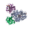
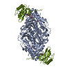
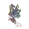
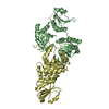
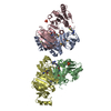
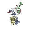
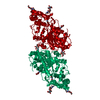
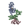
[日本語] English
- PDB-7p1h: Structure of the V. vulnificus ExoY-G-actin-profilin complex -

+
データを開く


IDまたはキーワード:

読み込み中...

-
基本情報

登録情報
データベース: PDB / ID: 7p1h
タイトルStructure of the V. vulnificus ExoY-G-actin-profilin complex
要素
  • Actin, cytoplasmic 1
  • Maltose/maltodextrin-binding periplasmic protein,RTX-toxin
  • Profilin-1
キーワードTOXIN / Bacterial toxin / G-actin / profilin
機能・相同性
機能・相同性情報


calcium- and calmodulin-responsive adenylate cyclase activity / synapse maturation / adenyl-nucleotide exchange factor activity / modification of postsynaptic actin cytoskeleton / positive regulation of norepinephrine uptake / negative regulation of actin filament bundle assembly / positive regulation of actin filament bundle assembly / negative regulation of actin filament polymerization / cellular response to cytochalasin B / bBAF complex ...calcium- and calmodulin-responsive adenylate cyclase activity / synapse maturation / adenyl-nucleotide exchange factor activity / modification of postsynaptic actin cytoskeleton / positive regulation of norepinephrine uptake / negative regulation of actin filament bundle assembly / positive regulation of actin filament bundle assembly / negative regulation of actin filament polymerization / cellular response to cytochalasin B / bBAF complex / regulation of actin filament polymerization / npBAF complex / nBAF complex / brahma complex / Signaling by ROBO receptors / regulation of transepithelial transport / Formation of annular gap junctions / morphogenesis of a polarized epithelium / Formation of the dystrophin-glycoprotein complex (DGC) / structural constituent of postsynaptic actin cytoskeleton / GBAF complex / Gap junction degradation / Folding of actin by CCT/TriC / regulation of G0 to G1 transition / protein localization to adherens junction / Cell-extracellular matrix interactions / dense body / positive regulation of ATP-dependent activity / postsynaptic actin cytoskeleton / Tat protein binding / Prefoldin mediated transfer of substrate to CCT/TriC / RSC-type complex / regulation of double-strand break repair / proline-rich region binding / regulation of nucleotide-excision repair / PCP/CE pathway / Adherens junctions interactions / RHOF GTPase cycle / adherens junction assembly / apical protein localization / positive regulation of ruffle assembly / Sensory processing of sound by outer hair cells of the cochlea / negative regulation of stress fiber assembly / Interaction between L1 and Ankyrins / tight junction / SWI/SNF complex / regulation of mitotic metaphase/anaphase transition / Sensory processing of sound by inner hair cells of the cochlea / host cell cytosol / positive regulation of T cell differentiation / apical junction complex / regulation of norepinephrine uptake / positive regulation of double-strand break repair / transporter regulator activity / detection of maltose stimulus / maintenance of blood-brain barrier / nitric-oxide synthase binding / maltose transport complex / cortical cytoskeleton / positive regulation of actin filament polymerization / NuA4 histone acetyltransferase complex / establishment or maintenance of cell polarity / positive regulation of stem cell population maintenance / Regulation of MITF-M-dependent genes involved in pigmentation / carbohydrate transport / Recycling pathway of L1 / brush border / regulation of G1/S transition of mitotic cell cycle / positive regulation of epithelial cell migration / actin monomer binding / kinesin binding / EPH-ephrin mediated repulsion of cells / negative regulation of cell differentiation / carbohydrate transmembrane transporter activity / maltose binding / RHO GTPases Activate WASPs and WAVEs / maltose transport / regulation of synaptic vesicle endocytosis / maltodextrin transmembrane transport / positive regulation of myoblast differentiation / RHO GTPases activate IQGAPs / regulation of protein localization to plasma membrane / positive regulation of double-strand break repair via homologous recombination / ATP-binding cassette (ABC) transporter complex, substrate-binding subunit-containing / phosphatidylinositol-4,5-bisphosphate binding / phosphotyrosine residue binding / EPHB-mediated forward signaling / cytoskeleton organization / substantia nigra development / ATP-binding cassette (ABC) transporter complex / axonogenesis / calyx of Held / nitric-oxide synthase regulator activity / cell chemotaxis / neural tube closure / Translocation of SLC2A4 (GLUT4) to the plasma membrane / FCGR3A-mediated phagocytosis / actin filament / adherens junction / positive regulation of cell differentiation
類似検索 - 分子機能
Yersinia/Haemophilus virulence surface antigen / Profilin1/2/3, vertebrate / Peptidase C58, YopT-type domain / Dermonecrotic/RTX toxin, membrane localization domain / Anthrax toxin, edema factor, central / Anthrax toxin, edema factor, C-terminal / Anthrax toxin, edema factor, central domain superfamily / Anthrax toxin LF subunit / Membrane Localization Domain / Profilin conserved site ...Yersinia/Haemophilus virulence surface antigen / Profilin1/2/3, vertebrate / Peptidase C58, YopT-type domain / Dermonecrotic/RTX toxin, membrane localization domain / Anthrax toxin, edema factor, central / Anthrax toxin, edema factor, C-terminal / Anthrax toxin, edema factor, central domain superfamily / Anthrax toxin LF subunit / Membrane Localization Domain / Profilin conserved site / Profilin signature. / Profilin / Profilin / : / Profilin / Profilin superfamily / CGT/MARTX, cysteine protease (CPD) domain / CGT/MARTX, cysteine protease (CPD) domain superfamily / Peptidase C80 family / CGT/MARTX cysteine protease (CPD) domain profile. / alpha/beta hydrolase fold / Maltose/Cyclodextrin ABC transporter, substrate-binding protein / Solute-binding family 1, conserved site / Bacterial extracellular solute-binding proteins, family 1 signature. / Bacterial extracellular solute-binding protein / Alpha/beta hydrolase fold-1 / Actins signature 1. / Actin, conserved site / Actins signature 2. / Bacterial extracellular solute-binding protein / Actin/actin-like conserved site / Actins and actin-related proteins signature. / Actin / Actin family / Actin / ATPase, nucleotide binding domain / Alpha/Beta hydrolase fold
類似検索 - ドメイン・相同性
ADENOSINE-5'-TRIPHOSPHATE / RTX-toxin / Profilin-1 / Maltose/maltodextrin-binding periplasmic protein / Actin, cytoplasmic 1
類似検索 - 構成要素
生物種Escherichia coli (大腸菌)
Vibrio vulnificus (バクテリア)
Homo sapiens (ヒト)
手法電子顕微鏡法 / 単粒子再構成法 / クライオ電子顕微鏡法 / 解像度: 3.9 Å
データ登録者Belyy, A. / Merino, F. / Raunser, S.
資金援助 ドイツ, 1件
組織認可番号
Max Planck Society ドイツ
引用ジャーナル: Nat Commun / : 2021
タイトル: Mechanism of actin-dependent activation of nucleotidyl cyclase toxins from bacterial human pathogens.
著者: Alexander Belyy / Felipe Merino / Undine Mechold / Stefan Raunser /
要旨: Bacterial human pathogens secrete initially inactive nucleotidyl cyclases that become potent enzymes by binding to actin inside eukaryotic host cells. The underlying molecular mechanism of this ...Bacterial human pathogens secrete initially inactive nucleotidyl cyclases that become potent enzymes by binding to actin inside eukaryotic host cells. The underlying molecular mechanism of this activation is, however, unclear. Here, we report structures of ExoY from Pseudomonas aeruginosa and Vibrio vulnificus bound to their corresponding activators F-actin and profilin-G-actin. The structures reveal that in contrast to the apo-state, two flexible regions become ordered and interact strongly with actin. The specific stabilization of these regions results in an allosteric stabilization of the nucleotide binding pocket and thereby to an activation of the enzyme. Differences in the sequence and conformation of the actin-binding regions are responsible for the selective binding to either F- or G-actin. Other nucleotidyl cyclase toxins that bind to calmodulin rather than actin undergo a similar disordered-to-ordered transition during activation, suggesting that the allosteric activation-by-stabilization mechanism of ExoY is conserved in these enzymes, albeit the different activator.
履歴
登録2021年7月1日登録サイト: PDBE / 処理サイト: PDBE
改定 1.02021年11月17日Provider: repository / タイプ: Initial release
改定 1.12021年12月1日Group: Database references / カテゴリ: citation / citation_author
Item: _citation.country / _citation.journal_abbrev ..._citation.country / _citation.journal_abbrev / _citation.journal_id_CSD / _citation.journal_id_ISSN / _citation.journal_volume / _citation.page_first / _citation.page_last / _citation.pdbx_database_id_PubMed / _citation.title / _citation.year / _citation_author.identifier_ORCID

-
構造の表示

ムービー
  • 登録構造単位
  • Jmolによる作画
  • ダウンロード
  • 単純化した表面モデル + あてはめた原子モデル
  • マップデータ: EMDB-13159
  • Jmolによる作画
  • ダウンロード
  • EMマップとの重ね合わせ
  • マップデータ: EMDB-13159
  • UCSF Chimeraによる作画
  • ダウンロード
ムービービューア
構造ビューア分子:
MolmilJmol/JSmol

ダウンロードとリンク

-
集合体

登録構造単位
A: Maltose/maltodextrin-binding periplasmic protein,RTX-toxin
B: Actin, cytoplasmic 1
P: Profilin-1
ヘテロ分子


分子量 (理論値)分子数
合計 (水以外)148,4885
ポリマ-147,9413
非ポリマー5472
00
1


  • 登録構造と同一
  • 登録者・ソフトウェアが定義した集合体
  • 根拠: microscopy
タイプ名称対称操作
identity operation1_5551
Buried area5970 Å2
ΔGint-34 kcal/mol
Surface area39120 Å2
手法PISA

-
要素

#1: タンパク質 Maltose/maltodextrin-binding periplasmic protein,RTX-toxin / MMBP / Maltodextrin-binding protein / Maltose-binding protein / MBP


分子量: 91467.539 Da / 分子数: 1 / 由来タイプ: 組換発現
由来: (組換発現) Escherichia coli (strain K12) (大腸菌), (組換発現) Vibrio vulnificus (バクテリア)
: K12 / 遺伝子: malE, b4034, JW3994 / 発現宿主: Escherichia coli (大腸菌) / 株 (発現宿主): BL21 DE3 RIPL / 参照: UniProt: P0AEX9, UniProt: A0A0F6NGV0
#2: タンパク質 Actin, cytoplasmic 1 / Beta-actin


分子量: 41402.242 Da / 分子数: 1 / Mutation: C272A / 由来タイプ: 組換発現 / 由来: (組換発現) Homo sapiens (ヒト) / 遺伝子: ACTB / 細胞株 (発現宿主): BTI-Tnao38 / 発現宿主: 
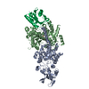
Trichoplusia ni (イラクサキンウワバ) / 参照: UniProt: P60709
#3: タンパク質 Profilin-1 / Epididymis tissue protein Li 184a / Profilin I


分子量: 15071.222 Da / 分子数: 1 / 由来タイプ: 組換発現 / 由来: (組換発現) Homo sapiens (ヒト) / 遺伝子: PFN1 / 発現宿主: Escherichia coli (大腸菌) / 参照: UniProt: P07737
#4: 化合物 ChemComp-CA / CALCIUM ION


分子量: 40.078 Da / 分子数: 1 / 由来タイプ: 合成 / : Ca
#5: 化合物 ChemComp-ATP / ADENOSINE-5'-TRIPHOSPHATE


分子量: 507.181 Da / 分子数: 1 / 由来タイプ: 合成 / : C10H16N5O13P3 / コメント: ATP, エネルギー貯蔵分子*YM
研究の焦点であるリガンドがあるかN

-
実験情報

-
実験

実験手法: 電子顕微鏡法
EM実験試料の集合状態: PARTICLE / 3次元再構成法: 単粒子再構成法

-
試料調製

構成要素名称: Structure of the V. vulnificus ExoY-G-actin-profilin complex
タイプ: COMPLEX / Entity ID: #1-#3 / 由来: MULTIPLE SOURCES
分子量実験値: NO
緩衝液pH: 8
試料包埋: NO / シャドウイング: NO / 染色: NO / 凍結: YES
試料支持グリッドの材料: COPPER / グリッドのサイズ: 300 divisions/in. / グリッドのタイプ: Quantifoil R2/1
急速凍結装置: FEI VITROBOT MARK III / 凍結剤: ETHANE / 湿度: 100 %

-
電子顕微鏡撮影

実験機器
モデル: Titan Krios / 画像提供: FEI Company
顕微鏡モデル: FEI TITAN KRIOS
電子銃電子線源: FIELD EMISSION GUN / 加速電圧: 300 kV / 照射モード: SPOT SCAN
電子レンズモード: BRIGHT FIELD / 最大 デフォーカス(公称値): 2500 nm / 最小 デフォーカス(公称値): 1200 nm
撮影平均露光時間: 2 sec. / 電子線照射量: 60 e/Å2 / 検出モード: SUPER-RESOLUTION / フィルム・検出器のモデル: GATAN K3 (6k x 4k) / 撮影したグリッド数: 1 / 実像数: 6879

-
解析

ソフトウェア名称: PHENIX / バージョン: 1.17.1_3660: / 分類: 精密化
EMソフトウェア
ID名称カテゴリ
1crYOLO粒子像選択
2EPU画像取得
4CTFFINDCTF補正
7ISOLDEモデルフィッティング
9SPHIRE初期オイラー角割当
10SPHIRE最終オイラー角割当
12SPHIRE3次元再構成
13PHENIXモデル精密化
CTF補正タイプ: PHASE FLIPPING AND AMPLITUDE CORRECTION
粒子像の選択選択した粒子像数: 1252333
対称性点対称性: C1 (非対称)
3次元再構成解像度: 3.9 Å / 解像度の算出法: FSC 0.143 CUT-OFF / 粒子像の数: 342081 / アルゴリズム: FOURIER SPACE / 対称性のタイプ: POINT
原子モデル構築プロトコル: FLEXIBLE FIT / 空間: REAL
原子モデル構築
IDPDB-IDPDB chain-ID 3D fitting-ID
16NBWA1
26NBWC1
拘束条件
Refine-IDタイプDev ideal
ELECTRON MICROSCOPYf_bond_d0.0057219
ELECTRON MICROSCOPYf_angle_d1.0229791
ELECTRON MICROSCOPYf_dihedral_angle_d23.2182673
ELECTRON MICROSCOPYf_chiral_restr0.0521086
ELECTRON MICROSCOPYf_plane_restr0.0061254

+
万見について

-
お知らせ

-
2022年2月9日: EMDBエントリの付随情報ファイルのフォーマットが新しくなりました

EMDBエントリの付随情報ファイルのフォーマットが新しくなりました

  • EMDBのヘッダファイルのバージョン3が、公式のフォーマットとなりました。
  • これまでは公式だったバージョン1.9は、アーカイブから削除されます。

関連情報:EMDBヘッダ

外部リンク:wwPDBはEMDBデータモデルのバージョン3へ移行します

-
2020年8月12日: 新型コロナ情報

新型コロナ情報

URL: https://pdbj.org/emnavi/covid19.php

新ページ: EM Navigatorに新型コロナウイルスの特設ページを開設しました。

関連情報:Covid-19情報 / 2020年3月5日: 新型コロナウイルスの構造データ

+
2020年3月5日: 新型コロナウイルスの構造データ

新型コロナウイルスの構造データ

関連情報:万見生物種 / 2020年8月12日: 新型コロナ情報

外部リンク:COVID-19特集ページ - PDBj / 今月の分子2020年2月:コロナウイルスプロテーアーゼ

+
2019年1月31日: EMDBのIDの桁数の変更

EMDBのIDの桁数の変更

  • EMDBエントリに付与されているアクセスコード(EMDB-ID)は4桁の数字(例、EMD-1234)でしたが、間もなく枯渇します。これまでの4桁のID番号は4桁のまま変更されませんが、4桁の数字を使い切った後に発行されるIDは5桁以上の数字(例、EMD-12345)になります。5桁のIDは2019年の春頃から発行される見通しです。
  • EM Navigator/万見では、接頭語「EMD-」は省略されています。

関連情報:Q: 「EMD」とは何ですか? / 万見/EM NavigatorにおけるID/アクセスコードの表記

外部リンク:EMDB Accession Codes are Changing Soon! / PDBjへお問い合わせ

+
2017年7月12日: PDB大規模アップデート

PDB大規模アップデート

  • 新バージョンのPDBx/mmCIF辞書形式に基づくデータがリリースされました。
  • 今回の更新はバージョン番号が4から5になる大規模なもので、全エントリデータの書き換えが行われる「Remediation」というアップデートに該当します。
  • このバージョンアップで、電子顕微鏡の実験手法に関する多くの項目の書式が改定されました(例:em_softwareなど)。
  • EM NavigatorとYorodumiでも、この改定に基づいた表示内容になります。

外部リンク:wwPDB Remediation / OneDepデータ基準に準拠した、より強化された内容のモデル構造ファイルが、PDBアーカイブで公開されました。

-
万見 (Yorodumi)

幾万の構造データを、幾万の視点から

  • 万見(Yorodumi)は、EMDB/PDB/SASBDBなどの構造データを閲覧するためのページです。
  • EM Navigatorの詳細ページの後継、Omokage検索のフロントエンドも兼ねています。

関連情報:EMDB / PDB / SASBDB / 3つのデータバンクの比較 / 万見検索 / 2016年8月31日: 新しいEM Navigatorと万見 / 万見文献 / Jmol/JSmol / 機能・相同性情報 / 新しいEM Navigatorと万見の変更点

他の情報も見る