Movie
Movie Controller


+ Open data

- Basic information
| Entry | Database: PDB / ID: 7p1h | ||||||
|---|---|---|---|---|---|---|---|
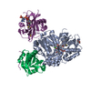
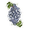
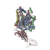
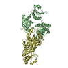
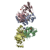
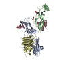
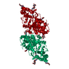
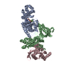
| Title | Structure of the V. vulnificus ExoY-G-actin-profilin complex | ||||||
 Components Components |
| ||||||
 Keywords Keywords | TOXIN / Bacterial toxin / G-actin / profilin | ||||||
| Function / homology |  Function and homology information Function and homology informationcalcium- and calmodulin-responsive adenylate cyclase activity / synapse maturation / adenyl-nucleotide exchange factor activity / modification of postsynaptic actin cytoskeleton / positive regulation of norepinephrine uptake / negative regulation of actin filament bundle assembly / positive regulation of actin filament bundle assembly / negative regulation of actin filament polymerization / bBAF complex / cellular response to cytochalasin B ...calcium- and calmodulin-responsive adenylate cyclase activity / synapse maturation / adenyl-nucleotide exchange factor activity / modification of postsynaptic actin cytoskeleton / positive regulation of norepinephrine uptake / negative regulation of actin filament bundle assembly / positive regulation of actin filament bundle assembly / negative regulation of actin filament polymerization / bBAF complex / cellular response to cytochalasin B / regulation of actin filament polymerization / npBAF complex / Signaling by ROBO receptors / nBAF complex / brahma complex / regulation of transepithelial transport / Formation of annular gap junctions / Formation of the dystrophin-glycoprotein complex (DGC) / morphogenesis of a polarized epithelium / structural constituent of postsynaptic actin cytoskeleton / Gap junction degradation / GBAF complex / Folding of actin by CCT/TriC / regulation of G0 to G1 transition / protein localization to adherens junction / Cell-extracellular matrix interactions / dense body / positive regulation of ATP-dependent activity / Tat protein binding / postsynaptic actin cytoskeleton / Prefoldin mediated transfer of substrate to CCT/TriC / RSC-type complex / proline-rich region binding / regulation of double-strand break repair / regulation of nucleotide-excision repair / PCP/CE pathway / Adherens junctions interactions / RHOF GTPase cycle / adherens junction assembly / apical protein localization / positive regulation of ruffle assembly / Sensory processing of sound by outer hair cells of the cochlea / negative regulation of stress fiber assembly / Interaction between L1 and Ankyrins / tight junction / SWI/SNF complex / regulation of mitotic metaphase/anaphase transition / Sensory processing of sound by inner hair cells of the cochlea / positive regulation of T cell differentiation / host cell cytosol / apical junction complex / positive regulation of double-strand break repair / maintenance of blood-brain barrier / detection of maltose stimulus / regulation of norepinephrine uptake / nitric-oxide synthase binding / transporter regulator activity / maltose transport complex / cortical cytoskeleton / establishment or maintenance of cell polarity / NuA4 histone acetyltransferase complex / positive regulation of stem cell population maintenance / positive regulation of actin filament polymerization / Recycling pathway of L1 / Regulation of MITF-M-dependent genes involved in pigmentation / carbohydrate transport / brush border / regulation of G1/S transition of mitotic cell cycle / positive regulation of epithelial cell migration / actin monomer binding / EPH-ephrin mediated repulsion of cells / negative regulation of cell differentiation / carbohydrate transmembrane transporter activity / maltose binding / kinesin binding / RHO GTPases Activate WASPs and WAVEs / maltose transport / regulation of synaptic vesicle endocytosis / maltodextrin transmembrane transport / positive regulation of myoblast differentiation / RHO GTPases activate IQGAPs / regulation of protein localization to plasma membrane / positive regulation of double-strand break repair via homologous recombination / ATP-binding cassette (ABC) transporter complex, substrate-binding subunit-containing / phosphatidylinositol-4,5-bisphosphate binding / phosphotyrosine residue binding / EPHB-mediated forward signaling / cytoskeleton organization / substantia nigra development / axonogenesis / ATP-binding cassette (ABC) transporter complex / calyx of Held / nitric-oxide synthase regulator activity / cell chemotaxis / FCGR3A-mediated phagocytosis / actin filament / neural tube closure / adherens junction / Translocation of SLC2A4 (GLUT4) to the plasma membrane / Regulation of endogenous retroelements by Piwi-interacting RNAs (piRNAs) Similarity search - Function | ||||||
| Biological species |  Vibrio vulnificus (bacteria) Vibrio vulnificus (bacteria) Homo sapiens (human) Homo sapiens (human) | ||||||
| Method | ELECTRON MICROSCOPY / single particle reconstruction / cryo EM / Resolution: 3.9 Å | ||||||
 Authors Authors | Belyy, A. / Merino, F. / Raunser, S. | ||||||
| Funding support |  Germany, 1items Germany, 1items
| ||||||
 Citation Citation | Journal: Nat Commun / Year: 2021 Title: Mechanism of actin-dependent activation of nucleotidyl cyclase toxins from bacterial human pathogens. Authors: Alexander Belyy / Felipe Merino / Undine Mechold / Stefan Raunser /  Abstract: Bacterial human pathogens secrete initially inactive nucleotidyl cyclases that become potent enzymes by binding to actin inside eukaryotic host cells. The underlying molecular mechanism of this ...Bacterial human pathogens secrete initially inactive nucleotidyl cyclases that become potent enzymes by binding to actin inside eukaryotic host cells. The underlying molecular mechanism of this activation is, however, unclear. Here, we report structures of ExoY from Pseudomonas aeruginosa and Vibrio vulnificus bound to their corresponding activators F-actin and profilin-G-actin. The structures reveal that in contrast to the apo-state, two flexible regions become ordered and interact strongly with actin. The specific stabilization of these regions results in an allosteric stabilization of the nucleotide binding pocket and thereby to an activation of the enzyme. Differences in the sequence and conformation of the actin-binding regions are responsible for the selective binding to either F- or G-actin. Other nucleotidyl cyclase toxins that bind to calmodulin rather than actin undergo a similar disordered-to-ordered transition during activation, suggesting that the allosteric activation-by-stabilization mechanism of ExoY is conserved in these enzymes, albeit the different activator. | ||||||
| History |
|
- Structure visualization
| Movie |
Movie viewer |
|---|---|
| Structure viewer | Molecule: MolmilJmol/JSmol |
- Downloads & links
-Download
| PDBx/mmCIF format | 7p1h.cif.gz | 308.2 KB | Display | PDBx/mmCIF format |
|---|---|---|---|---|
| PDB format | pdb7p1h.ent.gz | 252 KB | Display | PDB format |
| PDBx/mmJSON format | 7p1h.json.gz | Tree view | PDBx/mmJSON format | |
| Others |  Other downloads Other downloads |
-Validation report
| Summary document | 7p1h_validation.pdf.gz | 604.4 KB | Display | wwPDB validaton report |
|---|---|---|---|---|
| Full document | 7p1h_full_validation.pdf.gz | 616.8 KB | Display | |
| Data in XML | 7p1h_validation.xml.gz | 34.6 KB | Display | |
| Data in CIF | 7p1h_validation.cif.gz | 49.5 KB | Display | |
| Arichive directory | https://data.pdbj.org/pub/pdb/validation_reports/p1/7p1hftp://data.pdbj.org/pub/pdb/validation_reports/p1/7p1h | HTTPS FTP |
-Related structure data
| Related structure data |  13159MC  7p1gC M: map data used to model this data C: citing same article ( |
|---|---|
| Similar structure data |


-Links
 PDBj
PDBj


- Assembly
| Deposited unit | 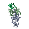
|
|---|---|
| 1 |
|
-Components
| #1: Protein | Mass: 91467.539 Da / Num. of mol.: 1 Source method: isolated from a genetically manipulated source Source: (gene. exp.) Vibrio vulnificus (bacteria)Strain: K12 / Gene: malE, b4034, JW3994 / Production host: |
|---|---|
| #2: Protein | Mass: 41402.242 Da / Num. of mol.: 1 / Mutation: C272A Source method: isolated from a genetically manipulated source Source: (gene. exp.) Homo sapiens (human) / Gene: ACTB / Cell line (production host): BTI-Tnao38 / Production host:  Trichoplusia ni (cabbage looper) / References: UniProt: P60709 Trichoplusia ni (cabbage looper) / References: UniProt: P60709 |
| #3: Protein | Mass: 15071.222 Da / Num. of mol.: 1 Source method: isolated from a genetically manipulated source Source: (gene. exp.) Homo sapiens (human) / Gene: PFN1 / Production host: |
| #4: Chemical | ChemComp-CA /   Mass: 40.078 Da / Num. of mol.: 1 / Source method: obtained synthetically / Formula: Ca Mass: 40.078 Da / Num. of mol.: 1 / Source method: obtained synthetically / Formula: Ca |
| #5: Chemical | ChemComp-ATP /   Mass: 507.181 Da / Num. of mol.: 1 / Source method: obtained synthetically / Formula: C10H16N5O13P3 / Comment: ATP, energy-carrying molecule*YM Mass: 507.181 Da / Num. of mol.: 1 / Source method: obtained synthetically / Formula: C10H16N5O13P3 / Comment: ATP, energy-carrying molecule*YM |
| Has ligand of interest | N |
-Experimental details
-Experiment
| Experiment | Method: ELECTRON MICROSCOPY |
|---|---|
| EM experiment | Aggregation state: PARTICLE / 3D reconstruction method: single particle reconstruction |
- Sample preparation
| Component | Name: Structure of the V. vulnificus ExoY-G-actin-profilin complex Type: COMPLEX / Entity ID: #1-#3 / Source: MULTIPLE SOURCES |
|---|---|
| Molecular weight | Experimental value: NO |
| Buffer solution | pH: 8 |
| Specimen | Embedding applied: NO / Shadowing applied: NO / Staining applied: NO / Vitrification applied: YES |
| Specimen support | Grid material: COPPER / Grid mesh size: 300 divisions/in. / Grid type: Quantifoil R2/1 |
| Vitrification | Instrument: FEI VITROBOT MARK III / Cryogen name: ETHANE / Humidity: 100 % |
- Electron microscopy imaging
| Experimental equipment |  Model: Titan Krios / Image courtesy: FEI Company |
|---|---|
| Microscopy | Model: FEI TITAN KRIOS |
| Electron gun | Electron source:  FIELD EMISSION GUN / Accelerating voltage: 300 kV / Illumination mode: SPOT SCAN FIELD EMISSION GUN / Accelerating voltage: 300 kV / Illumination mode: SPOT SCAN |
| Electron lens | Mode: BRIGHT FIELD / Nominal defocus max: 2500 nm / Nominal defocus min: 1200 nm |
| Image recording | Average exposure time: 2 sec. / Electron dose: 60 e/Å2 / Detector mode: SUPER-RESOLUTION / Film or detector model: GATAN K3 (6k x 4k) / Num. of grids imaged: 1 / Num. of real images: 6879 |
- Processing
| Software | Name: PHENIX / Version: 1.17.1_3660: / Classification: refinement | |||||||||||||||||||||||||||
|---|---|---|---|---|---|---|---|---|---|---|---|---|---|---|---|---|---|---|---|---|---|---|---|---|---|---|---|---|
| EM software |
| |||||||||||||||||||||||||||
| CTF correction | Type: PHASE FLIPPING AND AMPLITUDE CORRECTION | |||||||||||||||||||||||||||
| Particle selection | Num. of particles selected: 1252333 | |||||||||||||||||||||||||||
| Symmetry | Point symmetry: C1 (asymmetric) | |||||||||||||||||||||||||||
| 3D reconstruction | Resolution: 3.9 Å / Resolution method: FSC 0.143 CUT-OFF / Num. of particles: 342081 / Algorithm: FOURIER SPACE / Symmetry type: POINT | |||||||||||||||||||||||||||
| Atomic model building | Protocol: FLEXIBLE FIT / Space: REAL | |||||||||||||||||||||||||||
| Atomic model building |
| |||||||||||||||||||||||||||
| Refine LS restraints |
|