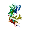
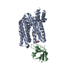
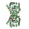
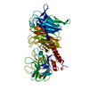
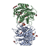
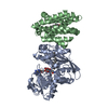
Journal: Elife / Year: 2021 Title: Cryo-EM structures of the caspase-activated protein XKR9 involved in apoptotic lipid scrambling. Authors: Monique S Straub / Carolina Alvadia / Marta Sawicka / Raimund Dutzler / Abstract: The exposure of the negatively charged lipid phosphatidylserine on the cell surface, catalyzed by lipid scramblases, is an important signal for the clearance of apoptotic cells by macrophages. The ...The exposure of the negatively charged lipid phosphatidylserine on the cell surface, catalyzed by lipid scramblases, is an important signal for the clearance of apoptotic cells by macrophages. The protein XKR9 is a member of a conserved family that has been associated with apoptotic lipid scrambling. Here, we describe structures of full-length and caspase-treated XKR9 from in complex with a synthetic nanobody determined by cryo-electron microscopy. The 43 kDa monomeric membrane protein can be divided into two structurally related repeats, each containing four membrane-spanning segments and a helix that is partly inserted into the lipid bilayer. In the full-length protein, the C-terminus interacts with a hydrophobic pocket located at the intracellular side acting as an inhibitor of protein function. Cleavage by caspase-3 at a specific site releases 16 residues of the C-terminus, thus making the pocket accessible to the cytoplasm. Collectively, the work has revealed the unknown architecture of the XKR family and has provided initial insight into its activation by caspases.
Mass: 792.075 Da / Num. of mol.: 1 / Source method: obtained synthetically / Formula: C42H82NO10P
Has ligand of interest
N
Has protein modification
Y
-
Experimental details
-
Experiment
Experiment
Method: ELECTRON MICROSCOPY
EM experiment
Aggregation state: PARTICLE / 3D reconstruction method: single particle reconstruction
-
Sample preparation
Component
ID
Name
Type
Entity ID
Parent-ID
Source
1
rXKR9withsybody
COMPLEX
#1-#2
0
RECOMBINANT
2
rXKR9
COMPLEX
#1
1
RECOMBINANT
3
Sybody
COMPLEX
#2
1
RECOMBINANT
Molecular weight
ID
Entity assembly-ID
Value (°)
Experimental value
1
1
0.06MDa
NO
2
1
0.043MDa
NO
3
1
0.017MDa
NO
Source (natural)
ID
Entity assembly-ID
Organism
Ncbi tax-ID
2
1
Rattus norvegicus (Norway rat)
10116
3
2
Rattus norvegicus (Norway rat)
10116
4
3
synthetic construct (others)
32630
Source (recombinant)
ID
Entity assembly-ID
Organism
Ncbi tax-ID
2
1
Homo sapiens (human)
9606
3
2
Homo sapiens (human)
9606
4
3
Escherichia coli MC1061 (bacteria)
1211845
Buffer solution
pH: 7.5
Buffer component
ID
Conc.
Name
Formula
Buffer-ID
1
200mM
sodiumchloride
NaCl
1
2
25mM
HEPES
HEPES
1
3
0.01 %
LaurylMaltoseNeopentylGlycol
LMNG
1
Specimen
Conc.: 1.5 mg/ml / Embedding applied: NO / Shadowing applied: NO / Staining applied: NO / Vitrification applied: YES / Details: This sample was monodisperse
In the structure databanks used in Yorodumi, some data are registered as the other names, "COVID-19 virus" and "2019-nCoV". Here are the details of the virus and the list of structure data.
Jan 31, 2019. EMDB accession codes are about to change! (news from PDBe EMDB page)
EMDB accession codes are about to change! (news from PDBe EMDB page)
The allocation of 4 digits for EMDB accession codes will soon come to an end. Whilst these codes will remain in use, new EMDB accession codes will include an additional digit and will expand incrementally as the available range of codes is exhausted. The current 4-digit format prefixed with “EMD-” (i.e. EMD-XXXX) will advance to a 5-digit format (i.e. EMD-XXXXX), and so on. It is currently estimated that the 4-digit codes will be depleted around Spring 2019, at which point the 5-digit format will come into force.
The EM Navigator/Yorodumi systems omit the EMD- prefix.
Related info.:Q: What is EMD? / ID/Accession-code notation in Yorodumi/EM Navigator
Yorodumi is a browser for structure data from EMDB, PDB, SASBDB, etc.
This page is also the successor to EM Navigator detail page, and also detail information page/front-end page for Omokage search.
The word "yorodu" (or yorozu) is an old Japanese word meaning "ten thousand". "mi" (miru) is to see.
Related info.:EMDB / PDB / SASBDB / Comparison of 3 databanks / Yorodumi Search / Aug 31, 2016. New EM Navigator & Yorodumi / Yorodumi Papers / Jmol/JSmol / Function and homology information / Changes in new EM Navigator and Yorodumi
 Movie
Movie Controller


 Open data
 Basic information
 Components
 Keywords
 Function and homology information
 Authors
 Switzerland, 2items
  Citation
 Structure visualization
 Downloads & links
 Other downloads


 PDBj
PDBj


 Assembly
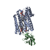
 Homo sapiens (human) / References: UniProt: Q5GH54

 Mass: 622.834 Da / Num. of mol.: 2 / Source method: obtained synthetically / Formula: C32H65NO8P / Comment: phospholipid*YM
 Mass: 792.075 Da / Num. of mol.: 1 / Source method: obtained synthetically / Formula: C42H82NO10P
 Sample preparation
 Electron microscopy imaging
 FIELD EMISSION GUN / Accelerating voltage: 300 kV / Illumination mode: FLOOD BEAM
 Processing