 Movie
Movie Controller

[English] 日本語
 Yorodumi
- PDB-7owe: Odinarchaeota Adenylate kinase (OdinAK) in complex with inhibitor Ap5a -
+ Open data

- Basic information
| Entry | Database: PDB / ID: 7owe | ||||||
|---|---|---|---|---|---|---|---|
| Title | Odinarchaeota Adenylate kinase (OdinAK) in complex with inhibitor Ap5a | ||||||
 Components Components | Adenylate kinase | ||||||
 Keywords Keywords | TRANSFERASE / Phosphotransferase / Odinarchaeota Adenylate kinase / AP5A | ||||||
| Function / homology | AAA domain / adenylate kinase / AMP kinase activity / phosphorylation / P-loop containing nucleoside triphosphate hydrolase / BIS(ADENOSINE)-5'-PENTAPHOSPHATE / Adenylate kinase Function and homology information Function and homology information | ||||||
| Biological species |  Candidatus Odinarchaeota archaeon LCB_4 (archaea) Candidatus Odinarchaeota archaeon LCB_4 (archaea) | ||||||
| Method |  X-RAY DIFFRACTION / SYNCHROTRON / MOLECULAR REPLACEMENT / Resolution: 2.75 Å X-RAY DIFFRACTION / SYNCHROTRON / MOLECULAR REPLACEMENT / Resolution: 2.75 Å | ||||||
 Authors Authors | Grundstrom, C. / Aberg-Zingmark, E. / Verma, A. / Wolf-Watz, M. / Sauer, U.H. / Sauer-Eriksson, A.E. | ||||||
| Funding support |  Sweden, 1items Sweden, 1items
| ||||||
 Citation Citation | Journal: Sci Adv / Year: 2022 Title: Insights into the evolution of enzymatic specificity and catalysis: From Asgard archaea to human adenylate kinases. Authors: Verma, A. / Aberg-Zingmark, E. / Sparrman, T. / Mushtaq, A.U. / Rogne, P. / Grundstrom, C. / Berntsson, R. / Sauer, U.H. / Backman, L. / Nam, K. / Sauer-Eriksson, E. / Wolf-Watz, M. | ||||||
| History |
|
- Structure visualization
| Structure viewer | Molecule: MolmilJmol/JSmol |
|---|
- Downloads & links
-Download
| PDBx/mmCIF format | 7owe.cif.gz | 244.2 KB | Display | PDBx/mmCIF format |
|---|---|---|---|---|
| PDB format | pdb7owe.ent.gz | 200.5 KB | Display | PDB format |
| PDBx/mmJSON format | 7owe.json.gz | Tree view | PDBx/mmJSON format | |
| Others |  Other downloads Other downloads |
-Validation report
| Arichive directory | https://data.pdbj.org/pub/pdb/validation_reports/ow/7oweftp://data.pdbj.org/pub/pdb/validation_reports/ow/7owe | HTTPS FTP |
|---|
-Related structure data
| Related structure data | 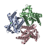 7owhC  7owjC 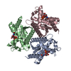 7owkC 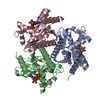 7owlC 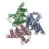 1ki9S S: Starting model for refinement C: citing same article ( |
|---|---|
| Similar structure data |
-Links
 PDBj
PDBj- Assembly
| Deposited unit | 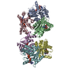
| ||||||||
|---|---|---|---|---|---|---|---|---|---|
| 1 | 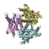
| ||||||||
| 2 | 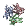
| ||||||||
| Unit cell |
|
-Components
| #1: Protein | Mass: 22883.516 Da / Num. of mol.: 6 Source method: isolated from a genetically manipulated source Source: (gene. exp.) Candidatus Odinarchaeota archaeon LCB_4 (archaea)Gene: adkA_1, OdinLCB4_00710 / Variant: Odinarchaeaota / Production host:  #2: Chemical | ChemComp-AP5 / 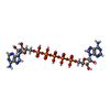  Mass: 916.367 Da / Num. of mol.: 6 / Source method: obtained synthetically / Formula: C20H29N10O22P5 / Feature type: SUBJECT OF INVESTIGATION Mass: 916.367 Da / Num. of mol.: 6 / Source method: obtained synthetically / Formula: C20H29N10O22P5 / Feature type: SUBJECT OF INVESTIGATIONHas ligand of interest | Y | |
|---|
-Experimental details
-Experiment
| Experiment | Method: X-RAY DIFFRACTION / Number of used crystals: 1 |
|---|
- Sample preparation
| Crystal | Density Matthews: 2.89 Å3/Da / Density % sol: 57.4 % |
|---|---|
| Crystal grow | Temperature: 298 K / Method: vapor diffusion, hanging drop / Details: Drop E7 PACT Screen: 0.2 M NaAc, 20% PEG 3550. |
-Data collection
| Diffraction | Mean temperature: 100 K / Serial crystal experiment: N |
|---|---|
| Diffraction source | Source: SYNCHROTRON / Site: PETRA III, EMBL c/o DESY  / Beamline: P13 (MX1) / Wavelength: 0.9762 Å / Beamline: P13 (MX1) / Wavelength: 0.9762 Å |
| Detector | Type: DECTRIS PILATUS 6M / Detector: PIXEL / Date: Sep 26, 2019 |
| Radiation | Protocol: SINGLE WAVELENGTH / Monochromatic (M) / Laue (L): M / Scattering type: x-ray |
| Radiation wavelength | Wavelength: 0.9762 Å / Relative weight: 1 |
| Reflection | Resolution: 2.75→71.6 Å / Num. obs: 42150 / % possible obs: 100 % / Redundancy: 13.1 % / CC1/2: 0.994 / Rmerge(I) obs: 0.135 / Rpim(I) all: 0.056 / Net I/σ(I): 9.3 |
| Reflection shell | Resolution: 2.75→2.85 Å / Redundancy: 12.5 % / Rmerge(I) obs: 1.98 / Mean I/σ(I) obs: 1.2 / Num. unique obs: 4139 / CC1/2: 0.626 / Rpim(I) all: 0.846 / % possible all: 100 |
- Processing
| Software |
| |||||||||||||||||||||||||||||||||||||||||||||||||||||||||||||||||||||||||||||||||||||
|---|---|---|---|---|---|---|---|---|---|---|---|---|---|---|---|---|---|---|---|---|---|---|---|---|---|---|---|---|---|---|---|---|---|---|---|---|---|---|---|---|---|---|---|---|---|---|---|---|---|---|---|---|---|---|---|---|---|---|---|---|---|---|---|---|---|---|---|---|---|---|---|---|---|---|---|---|---|---|---|---|---|---|---|---|---|---|
| Refinement | Method to determine structure: MOLECULAR REPLACEMENT Starting model: 1ki9 Resolution: 2.75→71.582 Å / SU ML: 0.44 / Cross valid method: THROUGHOUT / σ(F): 1.34 / Phase error: 33.2 / Stereochemistry target values: ML
| |||||||||||||||||||||||||||||||||||||||||||||||||||||||||||||||||||||||||||||||||||||
| Solvent computation | Shrinkage radii: 0.9 Å / VDW probe radii: 1.11 Å / Solvent model: FLAT BULK SOLVENT MODEL | |||||||||||||||||||||||||||||||||||||||||||||||||||||||||||||||||||||||||||||||||||||
| Displacement parameters | Biso max: 183.31 Å2 / Biso mean: 93.4972 Å2 / Biso min: 44.94 Å2 | |||||||||||||||||||||||||||||||||||||||||||||||||||||||||||||||||||||||||||||||||||||
| Refinement step | Cycle: final / Resolution: 2.75→71.582 Å
| |||||||||||||||||||||||||||||||||||||||||||||||||||||||||||||||||||||||||||||||||||||
| LS refinement shell | Refine-ID: X-RAY DIFFRACTION / Rfactor Rfree error: 0 / % reflection obs: 100 %
|
