 Movie
Movie Controller


+ Open data

- Basic information
| Entry | Database: PDB / ID: 1ki9 | ||||||
|---|---|---|---|---|---|---|---|
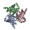
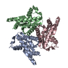
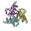
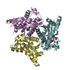
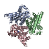
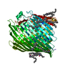
| Title | Adenylate kinase from Methanococcus thermolithotrophicus | ||||||
 Components Components | adenylate kinase | ||||||
 Keywords Keywords | SIGNALING PROTEIN / TRANSFERASE / kinase / phosphotransferase | ||||||
| Function / homology |  Function and homology information Function and homology information | ||||||
| Biological species |  Methanothermococcus thermolithotrophicus (archaea) Methanothermococcus thermolithotrophicus (archaea) | ||||||
| Method |  X-RAY DIFFRACTION / MOLECULAR REPLACEMENT / Resolution: 2.76 Å X-RAY DIFFRACTION / MOLECULAR REPLACEMENT / Resolution: 2.76 Å | ||||||
 Authors Authors | Criswell, A.R. / Konisky, J. / Phillips Jr., G.N. | ||||||
 Citation Citation | Journal: J.Mol.Biol. / Year: 2003 Title: Structures of thermophilic and mesophilic adenylate kinases from the genus Methanococcus Authors: Criswell, A.R. / Bae, E. / Stec, B. / Konisky, J. / Phillips Jr., G.N. | ||||||
| History |
|
- Structure visualization
| Structure viewer | Molecule: MolmilJmol/JSmol |
|---|
- Downloads & links
-Download
| PDBx/mmCIF format | 1ki9.cif.gz | 122.9 KB | Display | PDBx/mmCIF format |
|---|---|---|---|---|
| PDB format | pdb1ki9.ent.gz | 97.5 KB | Display | PDB format |
| PDBx/mmJSON format | 1ki9.json.gz | Tree view | PDBx/mmJSON format | |
| Others |  Other downloads Other downloads |
-Validation report
| Arichive directory | https://data.pdbj.org/pub/pdb/validation_reports/ki/1ki9ftp://data.pdbj.org/pub/pdb/validation_reports/ki/1ki9 | HTTPS FTP |
|---|
-Related structure data
| Related structure data | 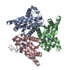 1khtC  1nksS C: citing same article ( S: Starting model for refinement |
|---|---|
| Similar structure data |


-Links
 PDBj
PDBj- Assembly
| Deposited unit | 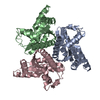
| ||||||||||
|---|---|---|---|---|---|---|---|---|---|---|---|
| 1 |
| ||||||||||
| 2 | 
| ||||||||||
| Unit cell |
|
-Components
| #1: Protein | Mass: 21486.764 Da / Num. of mol.: 3 Source method: isolated from a genetically manipulated source Source: (gene. exp.) Methanothermococcus thermolithotrophicus (archaea)Gene: adk / Plasmid: pET11a / Species (production host): Escherichia coli / Production host:  #2: Water | ChemComp-HOH / |  Mass: 18.015 Da / Num. of mol.: 160 / Source method: isolated from a natural source / Formula: H2O Mass: 18.015 Da / Num. of mol.: 160 / Source method: isolated from a natural source / Formula: H2O |
|---|
-Experimental details
-Experiment
| Experiment | Method: X-RAY DIFFRACTION / Number of used crystals: 1 |
|---|
- Sample preparation
| Crystal | Density Matthews: 2.8 Å3/Da / Density % sol: 55.7 % | ||||||||||||||||||||||||||||||||||||||||||
|---|---|---|---|---|---|---|---|---|---|---|---|---|---|---|---|---|---|---|---|---|---|---|---|---|---|---|---|---|---|---|---|---|---|---|---|---|---|---|---|---|---|---|---|
| Crystal grow | Temperature: 298 K / Method: vapor diffusion, hanging drop / pH: 7.5 Details: PEG 6000, MPD, HEPES, pH 7.5, VAPOR DIFFUSION, HANGING DROP at 298K | ||||||||||||||||||||||||||||||||||||||||||
| Crystal grow | *PLUS pH: 4.6 / Method: vapor diffusion, hanging drop | ||||||||||||||||||||||||||||||||||||||||||
| Components of the solutions | *PLUS
|
-Data collection
| Diffraction | Mean temperature: 100 K |
|---|---|
| Diffraction source | Source: ROTATING ANODE / Type: RIGAKU / Wavelength: 1.5418 Å |
| Detector | Type: RIGAKU RAXIS IV / Detector: IMAGE PLATE / Date: Feb 14, 1999 / Details: confocal |
| Radiation | Monochromator: mirrors / Protocol: SINGLE WAVELENGTH / Monochromatic (M) / Laue (L): M / Scattering type: x-ray |
| Radiation wavelength | Wavelength: 1.5418 Å / Relative weight: 1 |
| Reflection | Resolution: 2.5→50 Å / Num. all: 27043 / Num. obs: 25623 / % possible obs: 94.7 % / Biso Wilson estimate: 32.7 Å2 / Rmerge(I) obs: 0.068 |
| Reflection | *PLUS Highest resolution: 2.76 Å / % possible obs: 95.1 % / Rmerge(I) obs: 0.06 |
| Reflection shell | *PLUS Highest resolution: 2.76 Å / Lowest resolution: 2.95 Å / % possible obs: 94.4 % / Rmerge(I) obs: 0.175 |
- Processing
| Software |
| ||||||||||||||||||||||||||||||||||||
|---|---|---|---|---|---|---|---|---|---|---|---|---|---|---|---|---|---|---|---|---|---|---|---|---|---|---|---|---|---|---|---|---|---|---|---|---|---|
| Refinement | Method to determine structure: MOLECULAR REPLACEMENT Starting model: PDB entry 1NKS Resolution: 2.76→30.78 Å / Rfactor Rfree error: 0.007 / Data cutoff high absF: 157151.63 / Data cutoff low absF: 0 / Isotropic thermal model: RESTRAINED / Cross valid method: THROUGHOUT / σ(F): 0
| ||||||||||||||||||||||||||||||||||||
| Solvent computation | Solvent model: FLAT MODEL / Bsol: 32.3445 Å2 / ksol: 0.33371 e/Å3 | ||||||||||||||||||||||||||||||||||||
| Displacement parameters | Biso mean: 43 Å2
| ||||||||||||||||||||||||||||||||||||
| Refine analyze |
| ||||||||||||||||||||||||||||||||||||
| Refinement step | Cycle: LAST / Resolution: 2.76→30.78 Å
| ||||||||||||||||||||||||||||||||||||
| Refine LS restraints |
| ||||||||||||||||||||||||||||||||||||
| Xplor file |
| ||||||||||||||||||||||||||||||||||||
| Refinement | *PLUS | ||||||||||||||||||||||||||||||||||||
| Solvent computation | *PLUS | ||||||||||||||||||||||||||||||||||||
| Displacement parameters | *PLUS | ||||||||||||||||||||||||||||||||||||
| Refine LS restraints | *PLUS
|
