 Movie
Movie Controller

[English] 日本語
 Yorodumi
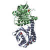
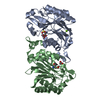
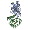
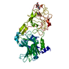
- PDB-7ovg: The C146A variant of an amidase from Pyrococcus horikoshii with b... -
+ Open data

- Basic information
| Entry | Database: PDB / ID: 7ovg | ||||||
|---|---|---|---|---|---|---|---|
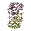
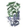
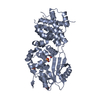
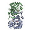
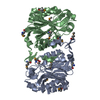
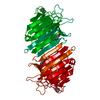
| Title | The C146A variant of an amidase from Pyrococcus horikoshii with bound acetamide | ||||||
 Components Components | CN hydrolase domain-containing protein | ||||||
 Keywords Keywords | HYDROLASE / Amidase / Nitrilase superfamily | ||||||
| Function / homology | : / : / Carbon-nitrogen hydrolase domain profile. / Carbon-nitrogen hydrolase superfamily / Carbon-nitrogen hydrolase / Carbon-nitrogen hydrolase / hydrolase activity, acting on carbon-nitrogen (but not peptide) bonds, in linear amides / ACETAMIDE / CN hydrolase domain-containing protein Function and homology information Function and homology information | ||||||
| Biological species |   Pyrococcus horikoshii (archaea) Pyrococcus horikoshii (archaea) | ||||||
| Method |  X-RAY DIFFRACTION / SYNCHROTRON / MOLECULAR REPLACEMENT / Resolution: 1.65 Å X-RAY DIFFRACTION / SYNCHROTRON / MOLECULAR REPLACEMENT / Resolution: 1.65 Å | ||||||
 Authors Authors | Su, S. / Makumire, S. / Sewell, B.T. | ||||||
| Funding support |  United Kingdom, 1items United Kingdom, 1items
| ||||||
 Citation Citation | Journal: J.Struct.Biol. / Year: 2022 Title: The structures of the C146A variant of the amidase from Pyrococcus horikoshii bound to glutaramide and acetamide suggest the basis of amide recognition. Authors: Makumire, S. / Su, S. / Weber, B.W. / Woodward, J.D. / Wangari Kimani, S. / Hunter, R. / Sewell, B.T. | ||||||
| History |
|
- Structure visualization
| Structure viewer | Molecule: MolmilJmol/JSmol |
|---|
- Downloads & links
-Download
| PDBx/mmCIF format | 7ovg.cif.gz | 121.2 KB | Display | PDBx/mmCIF format |
|---|---|---|---|---|
| PDB format | pdb7ovg.ent.gz | 92.2 KB | Display | PDB format |
| PDBx/mmJSON format | 7ovg.json.gz | Tree view | PDBx/mmJSON format | |
| Others |  Other downloads Other downloads |
-Validation report
| Arichive directory | https://data.pdbj.org/pub/pdb/validation_reports/ov/7ovgftp://data.pdbj.org/pub/pdb/validation_reports/ov/7ovg | HTTPS FTP |
|---|
-Related structure data
| Related structure data |  6ypaS S: Starting model for refinement |
|---|---|
| Similar structure data |
-Links
 PDBj
PDBj


- Assembly
| Deposited unit | 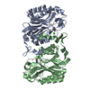
| ||||||||
|---|---|---|---|---|---|---|---|---|---|
| 1 |
| ||||||||
| Unit cell |
|
-Components
| #1: Protein | Mass: 29950.490 Da / Num. of mol.: 2 Source method: isolated from a genetically manipulated source Source: (gene. exp.) Pyrococcus horikoshii (strain ATCC 700860 / DSM 12428 / JCM 9974 / NBRC 100139 / OT-3) (archaea)Strain: ATCC 700860 / DSM 12428 / JCM 9974 / NBRC 100139 / OT-3 Gene: PH0642 / Production host:  #2: Chemical | 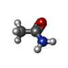  Mass: 59.067 Da / Num. of mol.: 2 / Source method: obtained synthetically / Formula: C2H5NO / Feature type: SUBJECT OF INVESTIGATION Mass: 59.067 Da / Num. of mol.: 2 / Source method: obtained synthetically / Formula: C2H5NO / Feature type: SUBJECT OF INVESTIGATION#3: Chemical |   Mass: 35.453 Da / Num. of mol.: 2 / Source method: obtained synthetically / Formula: Cl Mass: 35.453 Da / Num. of mol.: 2 / Source method: obtained synthetically / Formula: Cl#4: Water | ChemComp-HOH / |  Mass: 18.015 Da / Num. of mol.: 207 / Source method: isolated from a natural source / Formula: H2O Mass: 18.015 Da / Num. of mol.: 207 / Source method: isolated from a natural source / Formula: H2OHas ligand of interest | Y | |
|---|
-Experimental details
-Experiment
| Experiment | Method: X-RAY DIFFRACTION / Number of used crystals: 1 |
|---|
- Sample preparation
| Crystal | Density Matthews: 2.22 Å3/Da / Density % sol: 44.59 % |
|---|---|
| Crystal grow | Temperature: 293 K / Method: vapor diffusion, sitting drop Details: The crystallization condition is: 5mg/mL protein, 0.1M potassium chloride, 0.2M Sodium formate; 0.2M Ammonium acetate; 0.2M Sodium citrate tribasic dihydrate; 0.2M Sodium potassium tartrate ...Details: The crystallization condition is: 5mg/mL protein, 0.1M potassium chloride, 0.2M Sodium formate; 0.2M Ammonium acetate; 0.2M Sodium citrate tribasic dihydrate; 0.2M Sodium potassium tartrate tetrahydrate; 0.1M acetamide; Imidazole; MES monohydrate (acid); 25% v/v MPD; 25% PEG 1000; 25% w/v PEG 3350 |
-Data collection
| Diffraction | Mean temperature: 170 K / Serial crystal experiment: N |
|---|---|
| Diffraction source | Source: SYNCHROTRON / Site: Diamond / Beamline: I03 / Wavelength: 0.9763 Å |
| Detector | Type: DECTRIS EIGER2 XE 16M / Detector: PIXEL / Date: Sep 20, 2019 |
| Radiation | Protocol: SINGLE WAVELENGTH / Monochromatic (M) / Laue (L): M / Scattering type: x-ray |
| Radiation wavelength | Wavelength: 0.9763 Å / Relative weight: 1 |
| Reflection | Resolution: 1.65→43.86 Å / Num. obs: 53852 / % possible obs: 81.14 % / Redundancy: 3.4 % / Biso Wilson estimate: 22.57 Å2 / CC1/2: 0.996 / CC star: 0.999 / Rmerge(I) obs: 0.056 / Rpim(I) all: 0.035 / Rrim(I) all: 0.065 / Net I/σ(I): 11.32 |
| Reflection shell | Resolution: 1.65→1.709 Å / Redundancy: 3 % / Rmerge(I) obs: 0.3671 / Mean I/σ(I) obs: 2.39 / Num. unique obs: 2008 / CC1/2: 0.927 / CC star: 0.981 / Rpim(I) all: 0.2463 / Rrim(I) all: 0.4444 / % possible all: 30.28 |
- Processing
| Software |
| |||||||||||||||||||||||||||||||||||||||||||||||||||||||||||||||||||||||||||||||||||||||||||||||||||||||||
|---|---|---|---|---|---|---|---|---|---|---|---|---|---|---|---|---|---|---|---|---|---|---|---|---|---|---|---|---|---|---|---|---|---|---|---|---|---|---|---|---|---|---|---|---|---|---|---|---|---|---|---|---|---|---|---|---|---|---|---|---|---|---|---|---|---|---|---|---|---|---|---|---|---|---|---|---|---|---|---|---|---|---|---|---|---|---|---|---|---|---|---|---|---|---|---|---|---|---|---|---|---|---|---|---|---|---|
| Refinement | Method to determine structure: MOLECULAR REPLACEMENT Starting model: 6YPA Resolution: 1.65→43.85 Å / Cor.coef. Fo:Fc: 0.977 / Cor.coef. Fo:Fc free: 0.969 / SU B: 2.56 / SU ML: 0.078 / Cross valid method: THROUGHOUT / ESU R: 0.1 / ESU R Free: 0.097 / Stereochemistry target values: MAXIMUM LIKELIHOOD Details: HYDROGENS HAVE BEEN ADDED IN THE RIDING POSITIONS U VALUES : REFINED INDIVIDUALLY
| |||||||||||||||||||||||||||||||||||||||||||||||||||||||||||||||||||||||||||||||||||||||||||||||||||||||||
| Solvent computation | Ion probe radii: 0.8 Å / Shrinkage radii: 0.8 Å / VDW probe radii: 1.3 Å / Solvent model: MASK | |||||||||||||||||||||||||||||||||||||||||||||||||||||||||||||||||||||||||||||||||||||||||||||||||||||||||
| Displacement parameters | Biso mean: 29.106 Å2
| |||||||||||||||||||||||||||||||||||||||||||||||||||||||||||||||||||||||||||||||||||||||||||||||||||||||||
| Refinement step | Cycle: LAST / Resolution: 1.65→43.85 Å
| |||||||||||||||||||||||||||||||||||||||||||||||||||||||||||||||||||||||||||||||||||||||||||||||||||||||||
| Refine LS restraints |
| |||||||||||||||||||||||||||||||||||||||||||||||||||||||||||||||||||||||||||||||||||||||||||||||||||||||||
| LS refinement shell | Resolution: 1.65→1.693 Å / Total num. of bins used: 20
|
