 Movie
Movie Controller


+ Open data

- Basic information
| Entry | Database: PDB / ID: 7opk | ||||||
|---|---|---|---|---|---|---|---|
| Title | Crystal structure of C. thermophilum Xrn2 | ||||||
 Components Components | 5'-3' exoribonuclease | ||||||
 Keywords Keywords | RNA / degradation / metabolism / hydrolysis | ||||||
| Function / homology |  Function and homology information Function and homology information5'-3' RNA exonuclease activity / nuclear-transcribed mRNA catabolic process / DNA-templated transcription termination / mRNA processing / rRNA processing / Hydrolases; Acting on ester bonds; Exoribonucleases producing 5'-phosphomonoesters / RNA binding / metal ion binding / nucleus Similarity search - Function | ||||||
| Biological species |  Chaetomium thermophilum (fungus) Chaetomium thermophilum (fungus) | ||||||
| Method |  X-RAY DIFFRACTION / SYNCHROTRON / MOLECULAR REPLACEMENT / Resolution: 3 Å X-RAY DIFFRACTION / SYNCHROTRON / MOLECULAR REPLACEMENT / Resolution: 3 Å | ||||||
 Authors Authors | Overbeck, J.H. / Sprangers, R. | ||||||
 Citation Citation | Journal: Nat.Chem.Biol. / Year: 2022 Title: Observation of conformational changes that underlie the catalytic cycle of Xrn2. Authors: Overbeck, J.H. / Stelzig, D. / Fuchs, A.L. / Wurm, J.P. / Sprangers, R. | ||||||
| History |
|
- Structure visualization
| Structure viewer | Molecule: MolmilJmol/JSmol |
|---|
- Downloads & links
-Download
| PDBx/mmCIF format | 7opk.cif.gz | 332.9 KB | Display | PDBx/mmCIF format |
|---|---|---|---|---|
| PDB format | pdb7opk.ent.gz | 224.8 KB | Display | PDB format |
| PDBx/mmJSON format | 7opk.json.gz | Tree view | PDBx/mmJSON format | |
| Others |  Other downloads Other downloads |
-Validation report
| Arichive directory | https://data.pdbj.org/pub/pdb/validation_reports/op/7opkftp://data.pdbj.org/pub/pdb/validation_reports/op/7opk | HTTPS FTP |
|---|
-Related structure data
| Related structure data | 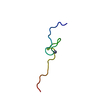 7pvmC 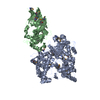 3fqdS S: Starting model for refinement C: citing same article ( |
|---|---|
| Similar structure data |
-Links
 PDBj
PDBj- Assembly
| Deposited unit | 
| ||||||||||||
|---|---|---|---|---|---|---|---|---|---|---|---|---|---|
| 1 |
| ||||||||||||
| Unit cell |
|
-Components
| #1: Protein | Mass: 100225.242 Da / Num. of mol.: 1 Source method: isolated from a genetically manipulated source Source: (gene. exp.) Chaetomium thermophilum (strain DSM 1495 / CBS 144.50 / IMI 039719) (fungus)Strain: DSM 1495 / CBS 144.50 / IMI 039719 / Gene: CTHT_0008830 / Production host:  References: UniProt: G0S058, Hydrolases; Acting on ester bonds; Exoribonucleases producing 5'-phosphomonoesters | ||||
|---|---|---|---|---|---|
| #2: Chemical |   Mass: 24.305 Da / Num. of mol.: 2 / Source method: obtained synthetically / Formula: Mg Mass: 24.305 Da / Num. of mol.: 2 / Source method: obtained synthetically / Formula: Mg#3: Water | ChemComp-HOH / |  Mass: 18.015 Da / Num. of mol.: 1 / Source method: isolated from a natural source / Formula: H2O Mass: 18.015 Da / Num. of mol.: 1 / Source method: isolated from a natural source / Formula: H2OHas ligand of interest | N | |
-Experimental details
-Experiment
| Experiment | Method: X-RAY DIFFRACTION / Number of used crystals: 1 |
|---|
- Sample preparation
| Crystal | Density Matthews: 3.94 Å3/Da / Density % sol: 68.76 % / Description: needles |
|---|---|
| Crystal grow | Temperature: 291 K / Method: vapor diffusion, sitting drop / Details: 0.2 M Magnesium formate, 20 %(w/v) PEG 3350 |
-Data collection
| Diffraction | Mean temperature: 100 K / Serial crystal experiment: N |
|---|---|
| Diffraction source | Source: SYNCHROTRON / Site: SLS  / Beamline: X10SA / Wavelength: 1 Å / Beamline: X10SA / Wavelength: 1 Å |
| Detector | Type: PSI PILATUS 6M / Detector: PIXEL / Date: May 2, 2017 |
| Radiation | Protocol: SINGLE WAVELENGTH / Monochromatic (M) / Laue (L): M / Scattering type: x-ray |
| Radiation wavelength | Wavelength: 1 Å / Relative weight: 1 |
| Reflection | Resolution: 3→50 Å / Num. obs: 31644 / % possible obs: 99.9 % / Redundancy: 20 % / Biso Wilson estimate: 63.06 Å2 / CC1/2: 0.996 / CC star: 0.999 / Rpim(I) all: 0.0679 / Rrim(I) all: 0.3085 / Net I/σ(I): 13.65 |
| Reflection shell | Resolution: 3→3.107 Å / Redundancy: 19.9 % / Mean I/σ(I) obs: 1.89 / Num. unique obs: 3130 / CC1/2: 0.786 / CC star: 0.938 / Rpim(I) all: 0.4547 / Rrim(I) all: 2.037 / % possible all: 99.94 |
- Processing
| Software |
| ||||||||||||||||||||||||||||||||||||||||||||||||||||||||||||||||||||||||||||||||||||
|---|---|---|---|---|---|---|---|---|---|---|---|---|---|---|---|---|---|---|---|---|---|---|---|---|---|---|---|---|---|---|---|---|---|---|---|---|---|---|---|---|---|---|---|---|---|---|---|---|---|---|---|---|---|---|---|---|---|---|---|---|---|---|---|---|---|---|---|---|---|---|---|---|---|---|---|---|---|---|---|---|---|---|---|---|---|
| Refinement | Method to determine structure: MOLECULAR REPLACEMENT Starting model: 3FQD Resolution: 3→49.69 Å / SU ML: 0.3746 / Cross valid method: FREE R-VALUE / σ(F): 1.37 / Phase error: 23.8028 Stereochemistry target values: GeoStd + Monomer Library + CDL v1.2
| ||||||||||||||||||||||||||||||||||||||||||||||||||||||||||||||||||||||||||||||||||||
| Solvent computation | Shrinkage radii: 0.9 Å / VDW probe radii: 1.11 Å / Solvent model: FLAT BULK SOLVENT MODEL | ||||||||||||||||||||||||||||||||||||||||||||||||||||||||||||||||||||||||||||||||||||
| Displacement parameters | Biso mean: 64.64 Å2 | ||||||||||||||||||||||||||||||||||||||||||||||||||||||||||||||||||||||||||||||||||||
| Refinement step | Cycle: LAST / Resolution: 3→49.69 Å
| ||||||||||||||||||||||||||||||||||||||||||||||||||||||||||||||||||||||||||||||||||||
| Refine LS restraints |
| ||||||||||||||||||||||||||||||||||||||||||||||||||||||||||||||||||||||||||||||||||||
| LS refinement shell |
| ||||||||||||||||||||||||||||||||||||||||||||||||||||||||||||||||||||||||||||||||||||
| Refinement TLS params. | Method: refined / Origin x: 88.7994438754 Å / Origin y: -26.9454813922 Å / Origin z: 0.189443296424 Å
| ||||||||||||||||||||||||||||||||||||||||||||||||||||||||||||||||||||||||||||||||||||
| Refinement TLS group | Selection details: all |
