 Movie
Movie Controller


+ Open data

- Basic information
| Entry | Database: PDB / ID: 7opj | ||||||
|---|---|---|---|---|---|---|---|
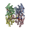
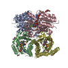
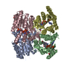
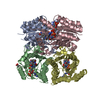
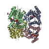
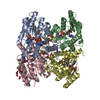
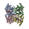
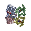
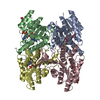
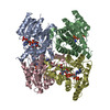
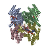
| Title | Trypanosoma brucei PTR1 (TbPTR1) in complex with pyrimethamine | ||||||
 Components Components | Pteridine reductase | ||||||
 Keywords Keywords | OXIDOREDUCTASE / Trypanosoma brucei / PTR1 / TbPTR1 / pyrimethamine | ||||||
| Function / homology |  Function and homology information Function and homology information | ||||||
| Biological species |  | ||||||
| Method |  X-RAY DIFFRACTION / SYNCHROTRON / MOLECULAR REPLACEMENT / Resolution: 1.34 Å X-RAY DIFFRACTION / SYNCHROTRON / MOLECULAR REPLACEMENT / Resolution: 1.34 Å | ||||||
 Authors Authors | Tassone, G. / Landi, G. / Pozzi, C. / Mangani, S. | ||||||
 Citation Citation | Journal: Pharmaceuticals / Year: 2021 Title: Evidence of Pyrimethamine and Cycloguanil Analogues as Dual Inhibitors of Trypanosoma brucei Pteridine Reductase and Dihydrofolate Reductase. Authors: Tassone, G. / Landi, G. / Linciano, P. / Francesconi, V. / Tonelli, M. / Tagliazucchi, L. / Costi, M.P. / Mangani, S. / Pozzi, C. | ||||||
| History |
|
- Structure visualization
| Structure viewer | Molecule: MolmilJmol/JSmol |
|---|
- Downloads & links
-Download
| PDBx/mmCIF format | 7opj.cif.gz | 428.8 KB | Display | PDBx/mmCIF format |
|---|---|---|---|---|
| PDB format | pdb7opj.ent.gz | 349.7 KB | Display | PDB format |
| PDBx/mmJSON format | 7opj.json.gz | Tree view | PDBx/mmJSON format | |
| Others |  Other downloads Other downloads |
-Validation report
| Arichive directory | https://data.pdbj.org/pub/pdb/validation_reports/op/7opjftp://data.pdbj.org/pub/pdb/validation_reports/op/7opj | HTTPS FTP |
|---|
-Related structure data
| Related structure data |  6tbxS S: Starting model for refinement |
|---|---|
| Similar structure data |


-Links
 PDBj
PDBj


- Assembly
| Deposited unit | 
| ||||||||
|---|---|---|---|---|---|---|---|---|---|
| 1 |
| ||||||||
| Unit cell |
|
-Components
-Protein , 1 types, 4 molecules ABCD
| #1: Protein | Mass: 30669.791 Da / Num. of mol.: 4 Source method: isolated from a genetically manipulated source Source: (gene. exp.)  |
|---|
-Non-polymers , 5 types, 1036 molecules 


| #2: Chemical | ChemComp-NAP /  Mass: 743.405 Da / Num. of mol.: 4 / Source method: obtained synthetically / Formula: C21H28N7O17P3 Mass: 743.405 Da / Num. of mol.: 4 / Source method: obtained synthetically / Formula: C21H28N7O17P3#3: Chemical | ChemComp-CP6 /  Mass: 248.711 Da / Num. of mol.: 4 / Source method: obtained synthetically / Formula: C12H13ClN4 / Feature type: SUBJECT OF INVESTIGATION / Comment: medication, antiparasitic*YM Mass: 248.711 Da / Num. of mol.: 4 / Source method: obtained synthetically / Formula: C12H13ClN4 / Feature type: SUBJECT OF INVESTIGATION / Comment: medication, antiparasitic*YM#4: Chemical | ChemComp-ACT /  Mass: 59.044 Da / Num. of mol.: 4 / Source method: obtained synthetically / Formula: C2H3O2 Mass: 59.044 Da / Num. of mol.: 4 / Source method: obtained synthetically / Formula: C2H3O2#5: Chemical | ChemComp-GOL / |  Mass: 92.094 Da / Num. of mol.: 1 / Source method: obtained synthetically / Formula: C3H8O3 Mass: 92.094 Da / Num. of mol.: 1 / Source method: obtained synthetically / Formula: C3H8O3#6: Water | ChemComp-HOH / | Mass: 18.015 Da / Num. of mol.: 1023 / Source method: isolated from a natural source / Formula: H2O |
|---|
-Details
| Has ligand of interest | Y |
|---|
-Experimental details
-Experiment
| Experiment | Method: X-RAY DIFFRACTION / Number of used crystals: 1 |
|---|
- Sample preparation
| Crystal | Density Matthews: 2.1 Å3/Da / Density % sol: 41.41 % |
|---|---|
| Crystal grow | Temperature: 294 K / Method: vapor diffusion, sitting drop / pH: 5 / Details: 2-2.5M sodium acetate, 0.1M sodium citrate, pH 5 |
-Data collection
| Diffraction | Mean temperature: 100 K / Serial crystal experiment: N |
|---|---|
| Diffraction source | Source: SYNCHROTRON / Site: Diamond  / Beamline: I03 / Wavelength: 0.9184 Å / Beamline: I03 / Wavelength: 0.9184 Å |
| Detector | Type: DECTRIS PILATUS3 6M / Detector: PIXEL / Date: Jul 29, 2015 |
| Radiation | Monochromator: Si(111) / Protocol: SINGLE WAVELENGTH / Monochromatic (M) / Laue (L): M / Scattering type: x-ray |
| Radiation wavelength | Wavelength: 0.9184 Å / Relative weight: 1 |
| Reflection | Resolution: 1.34→18.28 Å / Num. obs: 210248 / % possible obs: 93.9 % / Redundancy: 2.3 % / Biso Wilson estimate: 11.5 Å2 / CC1/2: 0.998 / Rmerge(I) obs: 0.056 / Rpim(I) all: 0.042 / Rrim(I) all: 0.071 / Net I/σ(I): 7.6 |
| Reflection shell | Resolution: 1.34→1.41 Å / Redundancy: 2.2 % / Rmerge(I) obs: 0.345 / Mean I/σ(I) obs: 2.3 / Num. unique obs: 30679 / CC1/2: 0.883 / Rpim(I) all: 0.27 / Rrim(I) all: 0.44 / % possible all: 94.2 |
- Processing
| Software |
| ||||||||||||||||||||||||||||||||||||||||||||||||||
|---|---|---|---|---|---|---|---|---|---|---|---|---|---|---|---|---|---|---|---|---|---|---|---|---|---|---|---|---|---|---|---|---|---|---|---|---|---|---|---|---|---|---|---|---|---|---|---|---|---|---|---|
| Refinement | Method to determine structure: MOLECULAR REPLACEMENT Starting model: 6TBX Resolution: 1.34→18.07 Å / Cor.coef. Fo:Fc: 0.981 / Cor.coef. Fo:Fc free: 0.97 / SU B: 1.992 / SU ML: 0.036 / Cross valid method: THROUGHOUT / σ(F): 0 / ESU R: 0.049 / ESU R Free: 0.051 / Stereochemistry target values: MAXIMUM LIKELIHOOD / Details: U VALUES : REFINED INDIVIDUALLY
| ||||||||||||||||||||||||||||||||||||||||||||||||||
| Solvent computation | Ion probe radii: 0.8 Å / Shrinkage radii: 0.8 Å / VDW probe radii: 1.2 Å / Solvent model: MASK | ||||||||||||||||||||||||||||||||||||||||||||||||||
| Displacement parameters | Biso max: 90.21 Å2 / Biso mean: 18.906 Å2 / Biso min: 6.78 Å2
| ||||||||||||||||||||||||||||||||||||||||||||||||||
| Refine analyze | Luzzati coordinate error obs: 0.1492 Å | ||||||||||||||||||||||||||||||||||||||||||||||||||
| Refinement step | Cycle: final / Resolution: 1.34→18.07 Å
| ||||||||||||||||||||||||||||||||||||||||||||||||||
| Refine LS restraints |
| ||||||||||||||||||||||||||||||||||||||||||||||||||
| LS refinement shell | Resolution: 1.34→1.375 Å / Rfactor Rfree error: 0 / Total num. of bins used: 20
|
