| Entry | Database: PDB / ID: 4wcd
|
|---|
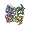
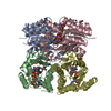
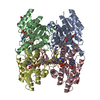
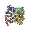
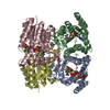
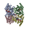
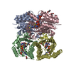
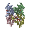
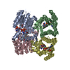
| Title | Trypanosoma brucei PTR1 in complex with inhibitor 10 |
|---|
 Components Components | Pteridine reductase |
|---|
 Keywords Keywords | OXIDOREDUCTASE / Thiadiazole derivatives inhibitors / TbPTR1 / Trypanosoma brucei PTR1 |
|---|
| Function / homology |  Function and homology information Function and homology information
Pteridine reductase / Short-chain dehydrogenase/reductase, conserved site / Short-chain dehydrogenases/reductases family signature. / Enoyl-(Acyl carrier protein) reductase / Short-chain dehydrogenase/reductase SDR / NAD(P)-binding Rossmann-like Domain / NAD(P)-binding domain superfamily / Rossmann fold / 3-Layer(aba) Sandwich / Alpha BetaSimilarity search - Domain/homology 5-(1H-benzotriazol-6-yl)-1,3,4-thiadiazol-2-amine / ACETATE ION / NADP NICOTINAMIDE-ADENINE-DINUCLEOTIDE PHOSPHATE / Pteridine reductaseSimilarity search - Component |
|---|
| Biological species |   Trypanosoma brucei brucei (eukaryote) Trypanosoma brucei brucei (eukaryote) |
|---|
| Method |  X-RAY DIFFRACTION / SYNCHROTRON / MOLECULAR REPLACEMENT / Resolution: 1.68 Å X-RAY DIFFRACTION / SYNCHROTRON / MOLECULAR REPLACEMENT / Resolution: 1.68 Å |
|---|
 Authors Authors | Mangani, S. / Di Pisa, F. / Pozzi, C. |
|---|
 Citation Citation | Journal: ACS Omega / Year: 2017
Title: Exploiting the 2-Amino-1,3,4-thiadiazole Scaffold To Inhibit Trypanosoma brucei Pteridine Reductase in Support of Early-Stage Drug Discovery.
Authors: Linciano, P. / Dawson, A. / Pohner, I. / Costa, D.M. / Sa, M.S. / Cordeiro-da-Silva, A. / Luciani, R. / Gul, S. / Witt, G. / Ellinger, B. / Kuzikov, M. / Gribbon, P. / Reinshagen, J. / Wolf, ...Authors: Linciano, P. / Dawson, A. / Pohner, I. / Costa, D.M. / Sa, M.S. / Cordeiro-da-Silva, A. / Luciani, R. / Gul, S. / Witt, G. / Ellinger, B. / Kuzikov, M. / Gribbon, P. / Reinshagen, J. / Wolf, M. / Behrens, B. / Hannaert, V. / Michels, P.A.M. / Nerini, E. / Pozzi, C. / di Pisa, F. / Landi, G. / Santarem, N. / Ferrari, S. / Saxena, P. / Lazzari, S. / Cannazza, G. / Freitas-Junior, L.H. / Moraes, C.B. / Pascoalino, B.S. / Alcantara, L.M. / Bertolacini, C.P. / Fontana, V. / Wittig, U. / Muller, W. / Wade, R.C. / Hunter, W.N. / Mangani, S. / Costantino, L. / Costi, M.P. |
|---|
| History | | Deposition | Sep 4, 2014 | Deposition site: RCSB / Processing site: PDBE |
|---|
| Revision 1.0 | Sep 30, 2015 | Provider: repository / Type: Initial release |
|---|
| Revision 1.1 | Sep 27, 2017 | Group: Database references / Structure summary / Category: audit_author / citation / citation_author
Item: _audit_author.identifier_ORCID / _citation.country ..._audit_author.identifier_ORCID / _citation.country / _citation.journal_abbrev / _citation.journal_id_CSD / _citation.journal_id_ISSN / _citation.journal_volume / _citation.page_first / _citation.page_last / _citation.pdbx_database_id_DOI / _citation.title / _citation.year |
|---|
| Revision 1.2 | Oct 18, 2017 | Group: Database references / Category: citation / citation_author
Item: _citation.journal_abbrev / _citation.pdbx_database_id_PubMed ..._citation.journal_abbrev / _citation.pdbx_database_id_PubMed / _citation.title / _citation_author.name |
|---|
| Revision 1.3 | Jan 10, 2024 | Group: Data collection / Database references / Refinement description
Category: chem_comp_atom / chem_comp_bond ...chem_comp_atom / chem_comp_bond / database_2 / pdbx_initial_refinement_model
Item: _database_2.pdbx_DOI / _database_2.pdbx_database_accession |
|---|
|
|---|
 Movie
Movie Controller


 Open data
 Basic information
 Structure visualization
 Downloads & links
 Other downloads


 PDBj
PDBj


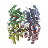
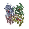
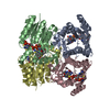
 Assembly
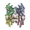


 Mass: 743.405 Da / Num. of mol.: 3 / Source method: obtained synthetically / Formula: C21H28N7O17P3
 Mass: 218.238 Da / Num. of mol.: 3 / Source method: obtained synthetically / Formula: C8H6N6S
 Mass: 59.044 Da / Num. of mol.: 3 / Source method: obtained synthetically / Formula: C2H3O2
 Mass: 92.094 Da / Num. of mol.: 1 / Source method: obtained synthetically / Formula: C3H8O3
 Sample preparation
 / Beamline: I04 / Wavelength: 0.97949 Å
 Processing