 Movie
Movie Controller


+ Open data

- Basic information
| Entry | Database: PDB / ID: 7omq | ||||||
|---|---|---|---|---|---|---|---|
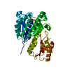
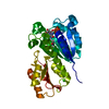
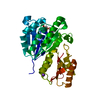
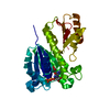
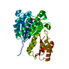
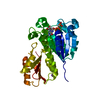
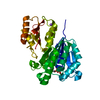
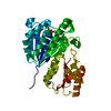
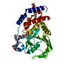
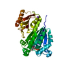
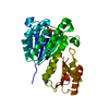
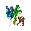
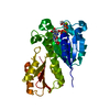
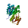
| Title | SaFtsZ complexed with GDPPCP and Mn2+ | ||||||
 Components Components | Cell division protein FtsZ | ||||||
 Keywords Keywords | CELL CYCLE / Cell division protein | ||||||
| Function / homology |  Function and homology information Function and homology informationcell septum / division septum assembly / FtsZ-dependent cytokinesis / cell division site / protein polymerization / GTPase activity / GTP binding / cytoplasm Similarity search - Function | ||||||
| Biological species |   Staphylococcus aureus (bacteria) Staphylococcus aureus (bacteria) | ||||||
| Method |  X-RAY DIFFRACTION / SYNCHROTRON / MOLECULAR REPLACEMENT / Resolution: 1.45 Å X-RAY DIFFRACTION / SYNCHROTRON / MOLECULAR REPLACEMENT / Resolution: 1.45 Å | ||||||
 Authors Authors | Fernandez-Tornero, C. / Ruiz, F.M. / Andreu, J.M. | ||||||
 Citation Citation | Journal: Plos Biol. / Year: 2022 Title: FtsZ filament structures in different nucleotide states reveal the mechanism of assembly dynamics. Authors: Ruiz, F.M. / Huecas, S. / Santos-Aledo, A. / Prim, E.A. / Andreu, J.M. / Fernandez-Tornero, C. | ||||||
| History |
|
- Structure visualization
| Structure viewer | Molecule: MolmilJmol/JSmol |
|---|
- Downloads & links
-Download
| PDBx/mmCIF format | 7omq.cif.gz | 222.5 KB | Display | PDBx/mmCIF format |
|---|---|---|---|---|
| PDB format | pdb7omq.ent.gz | 148 KB | Display | PDB format |
| PDBx/mmJSON format | 7omq.json.gz | Tree view | PDBx/mmJSON format | |
| Others |  Other downloads Other downloads |
-Validation report
| Arichive directory | https://data.pdbj.org/pub/pdb/validation_reports/om/7omqftp://data.pdbj.org/pub/pdb/validation_reports/om/7omq | HTTPS FTP |
|---|
-Related structure data
| Related structure data |  7ohhC  7ohkC  7ohlC 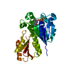 7ohnC  7oi2C  7ojaC  7ojbC  7ojcC  7ojdC  7ojzC  7omjC  7ompC  7on2C  7on3C  7on4C  6rvnS S: Starting model for refinement C: citing same article ( |
|---|---|
| Similar structure data |


-Links
 PDBj
PDBj


- Assembly
| Deposited unit | 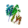
| ||||||||||||
|---|---|---|---|---|---|---|---|---|---|---|---|---|---|
| 1 |
| ||||||||||||
| Unit cell |
|
-Components
| #1: Protein | Mass: 31940.100 Da / Num. of mol.: 1 Source method: isolated from a genetically manipulated source Source: (gene. exp.) Staphylococcus aureus (bacteria) / Gene: ftsZ / Production host: | ||||
|---|---|---|---|---|---|
| #2: Chemical | ChemComp-GCP /   Mass: 521.208 Da / Num. of mol.: 1 / Source method: obtained synthetically / Formula: C11H18N5O13P3 / Feature type: SUBJECT OF INVESTIGATION / Comment: GMP-PCP, energy-carrying molecule analogue*YM Mass: 521.208 Da / Num. of mol.: 1 / Source method: obtained synthetically / Formula: C11H18N5O13P3 / Feature type: SUBJECT OF INVESTIGATION / Comment: GMP-PCP, energy-carrying molecule analogue*YM | ||||
| #3: Chemical | ChemComp-MN /   Mass: 54.938 Da / Num. of mol.: 1 / Source method: obtained synthetically / Formula: Mn Mass: 54.938 Da / Num. of mol.: 1 / Source method: obtained synthetically / Formula: Mn | ||||
| #4: Chemical |   Mass: 62.068 Da / Num. of mol.: 3 / Source method: obtained synthetically / Formula: C2H6O2 Mass: 62.068 Da / Num. of mol.: 3 / Source method: obtained synthetically / Formula: C2H6O2#5: Water | ChemComp-HOH / |  Mass: 18.015 Da / Num. of mol.: 204 / Source method: isolated from a natural source / Formula: H2O Mass: 18.015 Da / Num. of mol.: 204 / Source method: isolated from a natural source / Formula: H2OHas ligand of interest | Y | |
-Experimental details
-Experiment
| Experiment | Method: X-RAY DIFFRACTION / Number of used crystals: 1 |
|---|
- Sample preparation
| Crystal | Density Matthews: 2.37 Å3/Da / Density % sol: 48.15 % |
|---|---|
| Crystal grow | Temperature: 292 K / Method: vapor diffusion, sitting drop Details: 0.2 M lithium sulphate, 10% (v/v) ethylene glycol, 0.1 M Tris-HCl pH 8.5, and 22% polyethylene glycol 5000 monomethyl ether (+ 20 mM MnCl2 for soaking, 23h) |
-Data collection
| Diffraction | Mean temperature: 100 K / Serial crystal experiment: N |
|---|---|
| Diffraction source | Source: SYNCHROTRON / Site: ALBA  / Beamline: XALOC / Wavelength: 0.9793 Å / Beamline: XALOC / Wavelength: 0.9793 Å |
| Detector | Type: DECTRIS PILATUS 6M / Detector: PIXEL / Date: Mar 17, 2019 |
| Radiation | Protocol: SINGLE WAVELENGTH / Monochromatic (M) / Laue (L): M / Scattering type: x-ray |
| Radiation wavelength | Wavelength: 0.9793 Å / Relative weight: 1 |
| Reflection | Resolution: 1.45→40.98 Å / Num. obs: 52628 / % possible obs: 98.68 % / Redundancy: 4.4 % / Biso Wilson estimate: 20.77 Å2 / CC1/2: 0.998 / Net I/σ(I): 9.88 |
| Reflection shell | Resolution: 1.45→1.5 Å / Mean I/σ(I) obs: 1.11 / Num. unique obs: 5227 / CC1/2: 0.62 |
- Processing
| Software |
| |||||||||||||||||||||||||||||||||||||||||||||||||||||||||||||||||||||||||||||||||||||||||||||||||||||||||||||||||||||||||||||||||||||
|---|---|---|---|---|---|---|---|---|---|---|---|---|---|---|---|---|---|---|---|---|---|---|---|---|---|---|---|---|---|---|---|---|---|---|---|---|---|---|---|---|---|---|---|---|---|---|---|---|---|---|---|---|---|---|---|---|---|---|---|---|---|---|---|---|---|---|---|---|---|---|---|---|---|---|---|---|---|---|---|---|---|---|---|---|---|---|---|---|---|---|---|---|---|---|---|---|---|---|---|---|---|---|---|---|---|---|---|---|---|---|---|---|---|---|---|---|---|---|---|---|---|---|---|---|---|---|---|---|---|---|---|---|---|---|
| Refinement | Method to determine structure: MOLECULAR REPLACEMENT Starting model: 6RVN Resolution: 1.45→40.98 Å / SU ML: 0.1857 / Cross valid method: FREE R-VALUE / σ(F): 1.34 / Phase error: 22.5089 Stereochemistry target values: GeoStd + Monomer Library + CDL v1.2
| |||||||||||||||||||||||||||||||||||||||||||||||||||||||||||||||||||||||||||||||||||||||||||||||||||||||||||||||||||||||||||||||||||||
| Solvent computation | Shrinkage radii: 0.9 Å / VDW probe radii: 1.11 Å / Solvent model: FLAT BULK SOLVENT MODEL | |||||||||||||||||||||||||||||||||||||||||||||||||||||||||||||||||||||||||||||||||||||||||||||||||||||||||||||||||||||||||||||||||||||
| Displacement parameters | Biso mean: 31.63 Å2 | |||||||||||||||||||||||||||||||||||||||||||||||||||||||||||||||||||||||||||||||||||||||||||||||||||||||||||||||||||||||||||||||||||||
| Refinement step | Cycle: LAST / Resolution: 1.45→40.98 Å
| |||||||||||||||||||||||||||||||||||||||||||||||||||||||||||||||||||||||||||||||||||||||||||||||||||||||||||||||||||||||||||||||||||||
| Refine LS restraints |
| |||||||||||||||||||||||||||||||||||||||||||||||||||||||||||||||||||||||||||||||||||||||||||||||||||||||||||||||||||||||||||||||||||||
| LS refinement shell |
| |||||||||||||||||||||||||||||||||||||||||||||||||||||||||||||||||||||||||||||||||||||||||||||||||||||||||||||||||||||||||||||||||||||
| Refinement TLS params. | Method: refined / Refine-ID: X-RAY DIFFRACTION
| |||||||||||||||||||||||||||||||||||||||||||||||||||||||||||||||||||||||||||||||||||||||||||||||||||||||||||||||||||||||||||||||||||||
| Refinement TLS group | Refine-ID: X-RAY DIFFRACTION / Auth asym-ID: A / Label asym-ID: A
|
