 Movie
Movie Controller


+ Open data

- Basic information
| Entry | Database: PDB / ID: 7om0 | ||||||
|---|---|---|---|---|---|---|---|
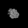
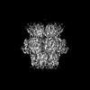
| Title | Structure of Primase-Helicase in SaPI5 | ||||||
 Components Components | DNA primase | ||||||
 Keywords Keywords | REPLICATION / Helicase / DNA binding / AMPPNP | ||||||
| Function / homology |  Function and homology information Function and homology information | ||||||
| Biological species |   Staphylococcus aureus (bacteria) Staphylococcus aureus (bacteria) | ||||||
| Method | ELECTRON MICROSCOPY / single particle reconstruction / cryo EM / Resolution: 3.1 Å | ||||||
 Authors Authors | Qiao, C.C. / Mir-Sanchis, I. | ||||||
| Funding support |  Sweden, 1items Sweden, 1items
| ||||||
 Citation Citation | Journal: Nucleic Acids Res / Year: 2022 Title: Staphylococcal self-loading helicases couple the staircase mechanism with inter domain high flexibility. Authors: Cuncun Qiao / Gianluca Debiasi-Anders / Ignacio Mir-Sanchis / Abstract: Replication is a crucial cellular process. Replicative helicases unwind DNA providing the template strand to the polymerase and promoting replication fork progression. Helicases are multi-domain ...Replication is a crucial cellular process. Replicative helicases unwind DNA providing the template strand to the polymerase and promoting replication fork progression. Helicases are multi-domain proteins which use an ATPase domain to couple ATP hydrolysis with translocation, however the role that the other domains might have during translocation remains elusive. Here, we studied the unexplored self-loading helicases called Reps, present in Staphylococcus aureus pathogenicity islands (SaPIs). Our cryoEM structures of the PriRep5 from SaPI5 (3.3 Å), the Rep1 from SaPI1 (3.9 Å) and Rep1-DNA complex (3.1Å) showed that in both Reps, the C-terminal domain (CTD) undergoes two distinct movements respect the ATPase domain. We experimentally demonstrate both in vitro and in vivo that SaPI-encoded Reps need key amino acids involved in the staircase mechanism of translocation. Additionally, we demonstrate that the CTD's presence is necessary for the maintenance of full ATPase and helicase activities. We speculate that this high interdomain flexibility couples Rep's activities as initiators and as helicases. | ||||||
| History |
|
- Structure visualization
| Structure viewer | Molecule: MolmilJmol/JSmol |
|---|
- Downloads & links
-Download
| PDBx/mmCIF format | 7om0.cif.gz | 502.2 KB | Display | PDBx/mmCIF format |
|---|---|---|---|---|
| PDB format | pdb7om0.ent.gz | 403 KB | Display | PDB format |
| PDBx/mmJSON format | 7om0.json.gz | Tree view | PDBx/mmJSON format | |
| Others |  Other downloads Other downloads |
-Validation report
| Arichive directory | https://data.pdbj.org/pub/pdb/validation_reports/om/7om0ftp://data.pdbj.org/pub/pdb/validation_reports/om/7om0 | HTTPS FTP |
|---|
-Related structure data
| Related structure data |  12982MC  7olaC 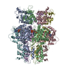 7pdsC C: citing same article ( M: map data used to model this data |
|---|---|
| Similar structure data | |
| EM raw data | EMPIAR-10881 (Title: Staphylococcal self-loading helicases couple the staircase mechanism with inter domain high flexibility Data size: 467.6 / Data #1: PriRep5-dsDNA [micrographs - multiframe]) |


-Links
 PDBj
PDBj


- Assembly
| Deposited unit | 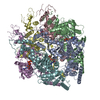
|
|---|---|
| 1 |
|
-Components
| #1: Protein | Mass: 93007.031 Da / Num. of mol.: 6 Source method: isolated from a genetically manipulated source Source: (gene. exp.) Staphylococcus aureus (bacteria)Gene: DQV53_04810, EQ90_04155, G6Y10_11370, HMPREF2819_07670 Production host: #2: Chemical | ChemComp-ANP /   Mass: 506.196 Da / Num. of mol.: 6 / Source method: obtained synthetically / Formula: C10H17N6O12P3 / Feature type: SUBJECT OF INVESTIGATION / Comment: AMP-PNP, energy-carrying molecule analogue*YM Mass: 506.196 Da / Num. of mol.: 6 / Source method: obtained synthetically / Formula: C10H17N6O12P3 / Feature type: SUBJECT OF INVESTIGATION / Comment: AMP-PNP, energy-carrying molecule analogue*YMHas ligand of interest | Y | Has protein modification | N | |
|---|
-Experimental details
-Experiment
| Experiment | Method: ELECTRON MICROSCOPY |
|---|---|
| EM experiment | Aggregation state: PARTICLE / 3D reconstruction method: single particle reconstruction |
- Sample preparation
| Component | Name: Primase-Helicase in SaPI5 / Type: ORGANELLE OR CELLULAR COMPONENT / Entity ID: #1 / Source: RECOMBINANT |
|---|---|
| Molecular weight | Value: 0.669 MDa / Experimental value: YES |
| Source (natural) | Organism: Staphylococcus aureus (bacteria) |
| Source (recombinant) | Organism: |
| Buffer solution | pH: 8 |
| Buffer component | Conc.: 200 mM / Name: Sodium Chloride / Formula: NaCl |
| Specimen | Conc.: 0.4 mg/ml / Embedding applied: NO / Shadowing applied: NO / Staining applied: NO / Vitrification applied: YES |
| Specimen support | Grid material: COPPER / Grid mesh size: 200 divisions/in. / Grid type: Quantifoil R2/2 |
| Vitrification | Instrument: FEI VITROBOT MARK IV / Cryogen name: ETHANE / Humidity: 100 % / Chamber temperature: 277 K |
- Electron microscopy imaging
| Experimental equipment |  Model: Titan Krios / Image courtesy: FEI Company |
|---|---|
| Microscopy | Model: FEI TITAN KRIOS |
| Electron gun | Electron source:  FIELD EMISSION GUN / Accelerating voltage: 300 kV / Illumination mode: SPOT SCAN FIELD EMISSION GUN / Accelerating voltage: 300 kV / Illumination mode: SPOT SCAN |
| Electron lens | Mode: BRIGHT FIELD / Nominal magnification: 165000 X / Cs: 2.7 mm |
| Specimen holder | Cryogen: NITROGEN / Specimen holder model: FEI TITAN KRIOS AUTOGRID HOLDER |
| Image recording | Average exposure time: 4 sec. / Electron dose: 1.16 e/Å2 / Detector mode: COUNTING / Film or detector model: GATAN K2 QUANTUM (4k x 4k) / Num. of real images: 4760 |
| Image scans | Movie frames/image: 40 |
- Processing
| EM software |
| |||||||||||||||||||||||||||||||||||||||||||||
|---|---|---|---|---|---|---|---|---|---|---|---|---|---|---|---|---|---|---|---|---|---|---|---|---|---|---|---|---|---|---|---|---|---|---|---|---|---|---|---|---|---|---|---|---|---|---|
| CTF correction | Details: CTF correction was performed by Gctf after motion correction. Type: PHASE FLIPPING AND AMPLITUDE CORRECTION | |||||||||||||||||||||||||||||||||||||||||||||
| Particle selection | Num. of particles selected: 724786 | |||||||||||||||||||||||||||||||||||||||||||||
| Symmetry | Point symmetry: C6 (6 fold cyclic) | |||||||||||||||||||||||||||||||||||||||||||||
| 3D reconstruction | Resolution: 3.1 Å / Resolution method: FSC 0.143 CUT-OFF / Num. of particles: 185358 / Symmetry type: POINT | |||||||||||||||||||||||||||||||||||||||||||||
| Atomic model building | Protocol: AB INITIO MODEL / Space: REAL / Target criteria: Correlation coefficient |
