 Movie
Movie Controller

[English] 日本語
 Yorodumi
- PDB-7ol9: Crystal structure of C-terminally truncated Bacillus subtilis nuc... -
+ Open data

- Basic information
| Entry | Database: PDB / ID: 7ol9 | |||||||||||||||
|---|---|---|---|---|---|---|---|---|---|---|---|---|---|---|---|---|
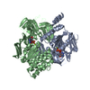
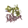
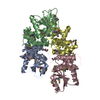
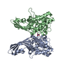
| Title | Crystal structure of C-terminally truncated Bacillus subtilis nucleoid occlusion protein (Noc) complexed to the Noc-binding site (NBS) | |||||||||||||||
 Components Components |
| |||||||||||||||
 Keywords Keywords | DNA BINDING PROTEIN / chromosome segregation / chromosome maintenance / protein-DNA recognition / DNA-binding protein | |||||||||||||||
| Function / homology |  Function and homology information Function and homology informationpositive regulation of sporulation resulting in formation of a cellular spore / division septum assembly / nucleoid / chromosome segregation / chromosome / DNA binding / cytoplasm Similarity search - Function | |||||||||||||||
| Biological species |  synthetic construct (others) | |||||||||||||||
| Method |  X-RAY DIFFRACTION / SYNCHROTRON / MOLECULAR REPLACEMENT / Resolution: 2.9 Å X-RAY DIFFRACTION / SYNCHROTRON / MOLECULAR REPLACEMENT / Resolution: 2.9 Å | |||||||||||||||
 Authors Authors | Jalal, A.S.B. / Lawson, D.M. / Le, T.B.K. | |||||||||||||||
| Funding support |  United Kingdom, 4items United Kingdom, 4items
| |||||||||||||||
 Citation Citation | Journal: J.Biol.Chem. / Year: 2023 Title: The CTP-binding domain is disengaged from the DNA-binding domain in a cocrystal structure of Bacillus subtilis Noc-DNA complex. Authors: Sukhoverkov, K.V. / Jalal, A.S.B. / Ault, J.R. / Sobott, F. / Lawson, D.M. / Le, T.B.K. #1: Journal: Biorxiv / Year: 2022Title: The CTP-binding domain is disengaged from the DNA-binding domain in a co-crystal structure of Bacillus subtilis Noc-DNA complex Authors: Sukhoverkov, K.V. / Jalal, A.S.B. / Lawson, D.M. / Le, T.B.K. | |||||||||||||||
| History |
|
- Structure visualization
| Structure viewer | Molecule: MolmilJmol/JSmol |
|---|
- Downloads & links
-Download
| PDBx/mmCIF format | 7ol9.cif.gz | 112.7 KB | Display | PDBx/mmCIF format |
|---|---|---|---|---|
| PDB format | pdb7ol9.ent.gz | 82.4 KB | Display | PDB format |
| PDBx/mmJSON format | 7ol9.json.gz | Tree view | PDBx/mmJSON format | |
| Others |  Other downloads Other downloads |
-Validation report
| Summary document | 7ol9_validation.pdf.gz | 451.6 KB | Display | wwPDB validaton report |
|---|---|---|---|---|
| Full document | 7ol9_full_validation.pdf.gz | 459.7 KB | Display | |
| Data in XML | 7ol9_validation.xml.gz | 15.9 KB | Display | |
| Data in CIF | 7ol9_validation.cif.gz | 20.9 KB | Display | |
| Arichive directory | https://data.pdbj.org/pub/pdb/validation_reports/ol/7ol9ftp://data.pdbj.org/pub/pdb/validation_reports/ol/7ol9 | HTTPS FTP |
-Related structure data
| Related structure data | 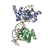 6y93S S: Starting model for refinement |
|---|---|
| Similar structure data |

-Links
 PDBj
PDBj


- Assembly
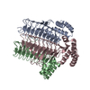
| Deposited unit | 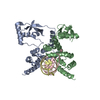
| ||||||||||||||||||||||||||||||||||||||||||||||||||||||||||||||||||||
|---|---|---|---|---|---|---|---|---|---|---|---|---|---|---|---|---|---|---|---|---|---|---|---|---|---|---|---|---|---|---|---|---|---|---|---|---|---|---|---|---|---|---|---|---|---|---|---|---|---|---|---|---|---|---|---|---|---|---|---|---|---|---|---|---|---|---|---|---|---|
| 1 |
| ||||||||||||||||||||||||||||||||||||||||||||||||||||||||||||||||||||
| Unit cell |
| ||||||||||||||||||||||||||||||||||||||||||||||||||||||||||||||||||||
| Noncrystallographic symmetry (NCS) | NCS domain:
NCS domain segments: Component-ID: _ / Refine code: _
NCS ensembles :
|
-Components
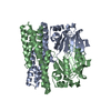
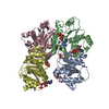
| #1: Protein | Mass: 29570.916 Da / Num. of mol.: 2 Source method: isolated from a genetically manipulated source Details: The expressed protein corresponds to residues 1-242 of UniProtKB - P37524. The C-terminal sequence KLAAALEHHHHHH is a nickel affinity tag from the pET21b expression plasmid. Source: (gene. exp.) Strain: 168 / Gene: noc, yyaA, BSU40990 / Plasmid: pET21b / Production host: #2: DNA chain | Mass: 4897.204 Da / Num. of mol.: 2 / Source method: obtained synthetically / Source: (synth.) synthetic construct (others) |
|---|
-Experimental details
-Experiment
| Experiment | Method: X-RAY DIFFRACTION / Number of used crystals: 1 |
|---|
- Sample preparation
| Crystal | Density Matthews: 2.52 Å3/Da / Density % sol: 51.3 % |
|---|---|
| Crystal grow | Temperature: 293 K / Method: vapor diffusion, sitting drop / pH: 8 / Details: Null |
-Data collection
| Diffraction | Mean temperature: 100 K / Serial crystal experiment: N | |||||||||||||||||||||
|---|---|---|---|---|---|---|---|---|---|---|---|---|---|---|---|---|---|---|---|---|---|---|
| Diffraction source | Source: SYNCHROTRON / Site: Diamond / Beamline: I04 / Wavelength: 0.9794 Å | |||||||||||||||||||||
| Detector | Type: DECTRIS EIGER2 XE 16M / Detector: PIXEL / Date: Sep 21, 2019 | |||||||||||||||||||||
| Radiation | Protocol: SINGLE WAVELENGTH / Monochromatic (M) / Laue (L): M / Scattering type: x-ray | |||||||||||||||||||||
| Radiation wavelength | Wavelength: 0.9794 Å / Relative weight: 1 | |||||||||||||||||||||
| Reflection | Resolution: 2.89→70.36 Å / Num. obs: 16242 / % possible obs: 100 % / Redundancy: 13.1 % / CC1/2: 0.999 / Rmerge(I) obs: 0.113 / Rpim(I) all: 0.032 / Rrim(I) all: 0.118 / Net I/σ(I): 11.6 | |||||||||||||||||||||
| Reflection shell | Diffraction-ID: 1 / % possible all: 100
|
- Processing
| Software |
| ||||||||||||||||||||||||||||||||||||||||||||||||||||||||||||
|---|---|---|---|---|---|---|---|---|---|---|---|---|---|---|---|---|---|---|---|---|---|---|---|---|---|---|---|---|---|---|---|---|---|---|---|---|---|---|---|---|---|---|---|---|---|---|---|---|---|---|---|---|---|---|---|---|---|---|---|---|---|
| Refinement | Method to determine structure: MOLECULAR REPLACEMENT Starting model: 6Y93 Resolution: 2.9→70.36 Å / Cor.coef. Fo:Fc: 0.924 / Cor.coef. Fo:Fc free: 0.934 / SU B: 20.896 / SU ML: 0.374 / Cross valid method: THROUGHOUT / σ(F): 0 / ESU R Free: 0.412 / Stereochemistry target values: MAXIMUM LIKELIHOOD Details: HYDROGENS HAVE BEEN ADDED IN THE RIDING POSITIONS U VALUES : REFINED INDIVIDUALLY
| ||||||||||||||||||||||||||||||||||||||||||||||||||||||||||||
| Solvent computation | Ion probe radii: 0.8 Å / Shrinkage radii: 0.8 Å / VDW probe radii: 1.2 Å / Solvent model: MASK | ||||||||||||||||||||||||||||||||||||||||||||||||||||||||||||
| Displacement parameters | Biso max: 201.63 Å2 / Biso mean: 109.446 Å2 / Biso min: 65.79 Å2
| ||||||||||||||||||||||||||||||||||||||||||||||||||||||||||||
| Refinement step | Cycle: final / Resolution: 2.9→70.36 Å
| ||||||||||||||||||||||||||||||||||||||||||||||||||||||||||||
| Refine LS restraints |
| ||||||||||||||||||||||||||||||||||||||||||||||||||||||||||||
| Refine LS restraints NCS | Refine-ID: X-RAY DIFFRACTION / Type: interatomic distance / Weight position: 0.05
| ||||||||||||||||||||||||||||||||||||||||||||||||||||||||||||
| LS refinement shell | Resolution: 2.9→2.975 Å / Rfactor Rfree error: 0 / Total num. of bins used: 20
|
