 Movie
Movie Controller


+ Open data

- Basic information
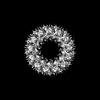
| Entry | Database: PDB / ID: 7ojf | ||||||
|---|---|---|---|---|---|---|---|
| Title | CRYO-EM STRUCTURE OF SLYB13-BAMA FROM ESCHERICHIA COLI. | ||||||
 Components Components | (Outer membrane ...) x 2 | ||||||
 Keywords Keywords | MEMBRANE PROTEIN / OUTER MEMBRANE CHAPERON / 2TM GLYCINE ZIPPER / OUTER MEMBRANE LIPOPROTEIN SLYB / LPS-LP BINDING PROTEIN | ||||||
| Function / homology |  Function and homology information Function and homology informationBam protein complex / outer membrane / Gram-negative-bacterium-type cell outer membrane assembly / protein insertion into membrane Similarity search - Function | ||||||
| Biological species |  | ||||||
| Method | ELECTRON MICROSCOPY / single particle reconstruction / cryo EM / Resolution: 3.9 Å | ||||||
 Authors Authors | Nguyen, V.S. / Remaut, H. | ||||||
 Citation Citation | Journal: Nature / Year: 2023 Title: SlyB encapsulates outer membrane proteins in stress-induced lipid nanodomains Authors: Janssens, A. / Nguyen, V.S. / Cecil, A.J. / Van der Verren, S.E. / Timmerman, E. / Deghelt, M. / Pak, A.J. / Collet, J.F. / Impens, F. / Remaut, H. | ||||||
| History |
|
- Structure visualization
| Structure viewer | Molecule: MolmilJmol/JSmol |
|---|
- Downloads & links
-Download
| PDBx/mmCIF format | 7ojf.cif.gz | 433.8 KB | Display | PDBx/mmCIF format |
|---|---|---|---|---|
| PDB format | pdb7ojf.ent.gz | Display | PDB format | |
| PDBx/mmJSON format | 7ojf.json.gz | Tree view | PDBx/mmJSON format | |
| Others |  Other downloads Other downloads |
-Validation report
| Arichive directory | https://data.pdbj.org/pub/pdb/validation_reports/oj/7ojfftp://data.pdbj.org/pub/pdb/validation_reports/oj/7ojf | HTTPS FTP |
|---|
-Related structure data
| Related structure data |  12949  7ojgC M: map data used to model this data C: citing same article ( |
|---|---|
| Similar structure data |


-Links
 PDBj
PDBj


- Assembly
| Deposited unit | 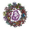
|
|---|---|
| 1 |
|
-Components
-Outer membrane ... , 2 types, 14 molecules ABCDEFGHIJKLMN
| #1: Protein | Mass: 15613.606 Da / Num. of mol.: 13 Source method: isolated from a genetically manipulated source Source: (gene. exp.) #2: Protein | | Mass: 90643.383 Da / Num. of mol.: 1 Source method: isolated from a genetically manipulated source Source: (gene. exp.) |
|---|
-Non-polymers , 4 types, 51 molecules 
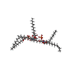
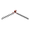

| #3: Chemical | ChemComp-PLM /  Mass: 256.424 Da / Num. of mol.: 13 / Source method: obtained synthetically / Formula: C16H32O2 Mass: 256.424 Da / Num. of mol.: 13 / Source method: obtained synthetically / Formula: C16H32O2#4: Chemical | ChemComp-L8Z / (  Mass: 2010.478 Da / Num. of mol.: 13 / Source method: obtained synthetically / Formula: C102H182N2O32P2 Mass: 2010.478 Da / Num. of mol.: 13 / Source method: obtained synthetically / Formula: C102H182N2O32P2#5: Chemical | ChemComp-LPP /  Mass: 648.891 Da / Num. of mol.: 12 / Source method: obtained synthetically / Formula: C35H69O8P / Comment: phospholipid*YM Mass: 648.891 Da / Num. of mol.: 12 / Source method: obtained synthetically / Formula: C35H69O8P / Comment: phospholipid*YM#6: Chemical | ChemComp-GOL /  Mass: 92.094 Da / Num. of mol.: 13 / Source method: obtained synthetically / Formula: C3H8O3 Mass: 92.094 Da / Num. of mol.: 13 / Source method: obtained synthetically / Formula: C3H8O3 |
|---|
-Details
| Has protein modification | Y |
|---|
-Experimental details
-Experiment
| Experiment | Method: ELECTRON MICROSCOPY |
|---|---|
| EM experiment | Aggregation state: PARTICLE / 3D reconstruction method: single particle reconstruction |
- Sample preparation
| Component | Name: Cryo-EM structure of SlyB13-BamA from Escherichia coli Type: COMPLEX Details: SlyB13-BamA was purified from overexpression of SlyB-TEV-HIS/BamA in E. coli using using 2-step Ni-IMAC purification. Entity ID: #1 / Source: RECOMBINANT | ||||||||||||||||||||
|---|---|---|---|---|---|---|---|---|---|---|---|---|---|---|---|---|---|---|---|---|---|
| Molecular weight | Value: 0.27 MDa / Experimental value: NO | ||||||||||||||||||||
| Source (natural) | Organism: | ||||||||||||||||||||
| Source (recombinant) | Organism: | ||||||||||||||||||||
| Buffer solution | pH: 8 | ||||||||||||||||||||
| Buffer component |
| ||||||||||||||||||||
| Specimen | Conc.: 0.04 mg/ml / Embedding applied: NO / Shadowing applied: NO / Staining applied: NO / Vitrification applied: YES | ||||||||||||||||||||
| Specimen support | Grid material: COPPER / Grid type: Quantifoil R2/1 | ||||||||||||||||||||
| Vitrification | Instrument: GATAN CRYOPLUNGE 3 / Cryogen name: ETHANE / Humidity: 90 % / Details: Back-blotting for 4 seconds before plunging |
- Electron microscopy imaging
| Microscopy | Model: JEOL CRYO ARM 300 |
|---|---|
| Electron gun | Electron source:  FIELD EMISSION GUN / Accelerating voltage: 300 kV / Illumination mode: OTHER FIELD EMISSION GUN / Accelerating voltage: 300 kV / Illumination mode: OTHER |
| Electron lens | Mode: BRIGHT FIELD / Nominal magnification: 60000 X / Nominal defocus max: 2500 nm / Nominal defocus min: 600 nm / Cs: 2.55 mm / Alignment procedure: COMA FREE |
| Specimen holder | Cryogen: NITROGEN / Specimen holder model: JEOL CRYOSPECPORTER |
| Image recording | Average exposure time: 3 sec. / Electron dose: 61 e/Å2 / Film or detector model: GATAN K3 (6k x 4k) / Num. of grids imaged: 1 / Num. of real images: 7352 |
| Image scans | Num. digital images: 7352 |
- Processing
| EM software |
| ||||||||||||||||||||||||||||||||||||||||||||||||||
|---|---|---|---|---|---|---|---|---|---|---|---|---|---|---|---|---|---|---|---|---|---|---|---|---|---|---|---|---|---|---|---|---|---|---|---|---|---|---|---|---|---|---|---|---|---|---|---|---|---|---|---|
| CTF correction | Type: NONE | ||||||||||||||||||||||||||||||||||||||||||||||||||
| 3D reconstruction | Resolution: 3.9 Å / Resolution method: FSC 0.143 CUT-OFF / Num. of particles: 73756 / Symmetry type: POINT | ||||||||||||||||||||||||||||||||||||||||||||||||||
| Atomic model building | B value: 56 / Protocol: OTHER / Space: REAL | ||||||||||||||||||||||||||||||||||||||||||||||||||
| Refinement | Highest resolution: 3.9 Å | ||||||||||||||||||||||||||||||||||||||||||||||||||
| Displacement parameters | Biso mean: 60.14 Å2 |
