Movie
Movie Controller

[English] 日本語
 Yorodumi
- EMDB-12946: Cryo-EM reconstruction of dodecameric SlyB from Escherichia coli K12 -
+ Open data

- Basic information
| Entry |  | |||||||||||||||
|---|---|---|---|---|---|---|---|---|---|---|---|---|---|---|---|---|
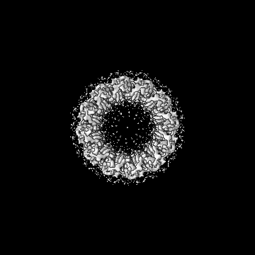
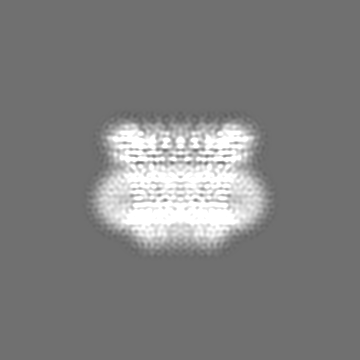
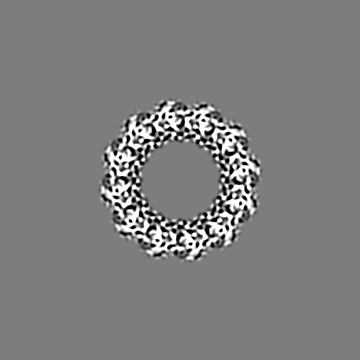
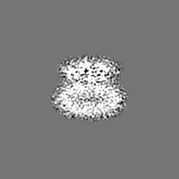
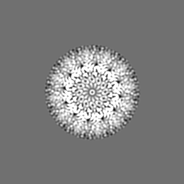
| Title | Cryo-EM reconstruction of dodecameric SlyB from Escherichia coli K12 | |||||||||||||||
 Map data Map data | SlyB12 | |||||||||||||||
 Sample Sample |
| |||||||||||||||
 Keywords Keywords | Outer membrane chaperon / 2TM glycine zipper / outer membrane lipoprotein SlyB / LPS-LP binding protein / MEMBRANE PROTEIN | |||||||||||||||
| Biological species |  | |||||||||||||||
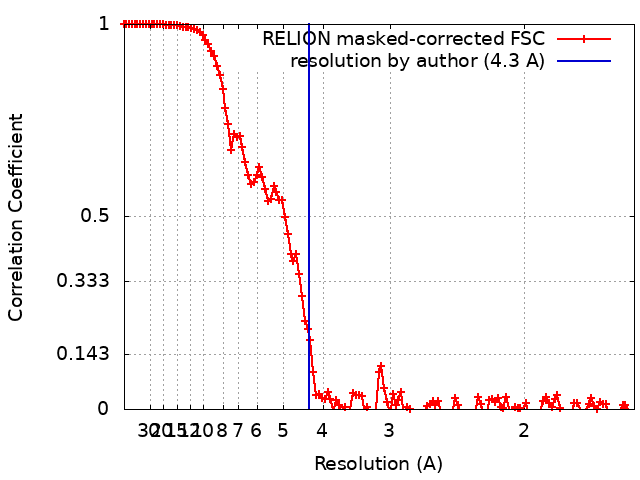
| Method | single particle reconstruction / cryo EM / Resolution: 4.3 Å | |||||||||||||||
 Authors Authors | Nguyen VS / Remaut H | |||||||||||||||
| Funding support |  Belgium, 4 items Belgium, 4 items
| |||||||||||||||
 Citation Citation | Journal: Nature / Year: 2023 Title: SlyB encapsulates outer membrane proteins in stress-induced lipid nanodomains Authors: Janssens A / Nguyen VS / Cecil AJ / Van der Verren SE / Timmerman E / Deghelt M / Pak AJ / Collet JF / Impens F / Remaut H | |||||||||||||||
| History |
|
- Structure visualization
| Supplemental images |
|---|

- Downloads & links
-EMDB archive
| Map data | emd_12946.map.gz | 18.6 MB |  EMDB map data format EMDB map data format | |
|---|---|---|---|---|
| Header (meta data) | emd-12946-v30.xmlemd-12946.xml | 12.5 KB 12.5 KB | Display Display | EMDB header |
| FSC (resolution estimation) | emd_12946_fsc.xml | 12.9 KB | Display | FSC data file |
| Images | 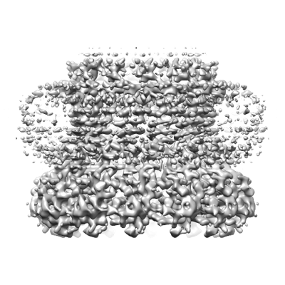 emd_12946.png emd_12946.png | 171.1 KB | ||
| Filedesc metadata | emd-12946.cif.gz | 5 KB | ||
| Archive directory |  http://ftp.pdbj.org/pub/emdb/structures/EMD-12946ftp://ftp.pdbj.org/pub/emdb/structures/EMD-12946 http://ftp.pdbj.org/pub/emdb/structures/EMD-12946ftp://ftp.pdbj.org/pub/emdb/structures/EMD-12946 | HTTPS FTP |
-Related structure data


-Links
| EMDB pages | EMDB (EBI/PDBe) / EMDataResource |
|---|
-Map
| File | Download / File: emd_12946.map.gz / Format: CCP4 / Size: 178 MB / Type: IMAGE STORED AS FLOATING POINT NUMBER (4 BYTES) | ||||||||||||||||||||||||||||||||||||
|---|---|---|---|---|---|---|---|---|---|---|---|---|---|---|---|---|---|---|---|---|---|---|---|---|---|---|---|---|---|---|---|---|---|---|---|---|---|
| Annotation | SlyB12 | ||||||||||||||||||||||||||||||||||||
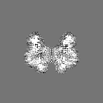
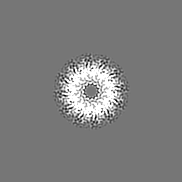
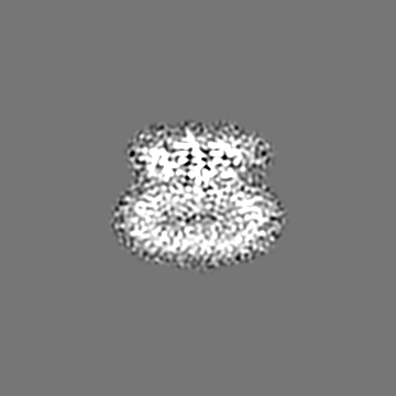
| Projections & slices | Image control
Images are generated by Spider. | ||||||||||||||||||||||||||||||||||||
| Voxel size | X=Y=Z: 0.784 Å | ||||||||||||||||||||||||||||||||||||
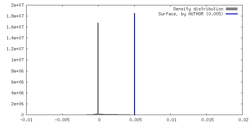
| Density |
| ||||||||||||||||||||||||||||||||||||
| Symmetry | Space group: 1 | ||||||||||||||||||||||||||||||||||||
| Details | EMDB XML:
|
 Z (Sec.)
 Y (Row.)
 X (Col.)


-Supplemental data
- Sample components
-Entire : Outer membrane lipoprotein SlyB
| Entire | Name: Outer membrane lipoprotein SlyB |
|---|---|
| Components |
|
-Supramolecule #1: Outer membrane lipoprotein SlyB
| Supramolecule | Name: Outer membrane lipoprotein SlyB / type: complex / ID: 1 / Parent: 0 / Macromolecule list: all Details: Dodecameric SlyB was purified from overexpression of SlyB-TEV-HIS in E. coli using using 2-step Ni-IMAC purification. |
|---|---|
| Source (natural) | Organism: |
| Molecular weight | Theoretical: 220 KDa |
-Macromolecule #1: Outer membrane lipoprotein SlyB
| Macromolecule | Name: Outer membrane lipoprotein SlyB / type: protein_or_peptide / ID: 1 / Enantiomer: LEVO |
|---|---|
| Source (natural) | Organism: |
| Recombinant expression | Organism: |
| Sequence | String: (PLM)CVNNDTLSG DVYTASEAKQ VQNVSYGTIV NVRPVQIQGG DDSNVIGAIG GAVLGGFLGN TVGGGTGRSL ATAAGAVAGG VAGQGVQSAM NKTQGVELEI RKDDGNTIMV VQKQGNTRFS PGQRVVLASN GSQVTVSPRG GGENLYFQ |
-Experimental details
-Structure determination
| Method | cryo EM |
|---|---|
 Processing Processing | single particle reconstruction |
| Aggregation state | particle |
-Sample preparation
| Concentration | 0.04 mg/mL | ||||||||||||
|---|---|---|---|---|---|---|---|---|---|---|---|---|---|
| Buffer | pH: 8 Component:
| ||||||||||||
| Grid | Model: Quantifoil R2/1 / Material: COPPER / Support film - Material: GRAPHENE OXIDE / Support film - topology: HOLEY / Pretreatment - Type: GLOW DISCHARGE / Pretreatment - Time: 60 sec. | ||||||||||||
| Vitrification | Cryogen name: ETHANE / Chamber humidity: 90 % / Instrument: GATAN CRYOPLUNGE 3 / Details: Back-blotting for 4 seconds before plunging. |
- Electron microscopy
| Microscope | JEOL CRYO ARM 300 |
|---|---|
| Image recording | Film or detector model: GATAN K3 (6k x 4k) / Number grids imaged: 1 / Number real images: 6352 / Average exposure time: 3.0 sec. / Average electron dose: 61.0 e/Å2 |
| Electron beam | Acceleration voltage: 300 kV / Electron source:  FIELD EMISSION GUN FIELD EMISSION GUN |
| Electron optics | Illumination mode: OTHER / Imaging mode: BRIGHT FIELD / Cs: 2.55 mm / Nominal defocus max: 2.5 µm / Nominal defocus min: 0.6 µm / Nominal magnification: 60000 |
| Sample stage | Specimen holder model: JEOL CRYOSPECPORTER / Cooling holder cryogen: NITROGEN |