 Movie
Movie Controller

[English] 日本語
 Yorodumi
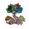
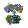
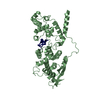
- PDB-7oi3: Cryo-EM structure of the Cetacean morbillivirus nucleoprotein-RNA... -
+ Open data

- Basic information
| Entry | Database: PDB / ID: 7oi3 | ||||||
|---|---|---|---|---|---|---|---|
| Title | Cryo-EM structure of the Cetacean morbillivirus nucleoprotein-RNA complex | ||||||
 Components Components |
| ||||||
 Keywords Keywords | VIRAL PROTEIN / Cetacean morbillivirus / nucleocapsid / RNA virus replication / cryo-electron microscopy / helical reconstruction / single particle analysis | ||||||
| Function / homology |  Function and homology information Function and homology informationhelical viral capsid / viral nucleocapsid / host cell cytoplasm / ribonucleoprotein complex / structural molecule activity / RNA binding Similarity search - Function | ||||||
| Biological species |  Morbillivirus sp. Morbillivirus sp. | ||||||
| Method | ELECTRON MICROSCOPY / helical reconstruction / cryo EM / Resolution: 4 Å | ||||||
 Authors Authors | Zinzula, L. / Beck, F. / Klumpe, S. / Bohn, S. / Pfeifer, G. / Bollschweiler, D. / Nagy, I. / Plitzko, J.M. / Baumeister, W. | ||||||
 Citation Citation | Journal: J Struct Biol / Year: 2021 Title: Cryo-EM structure of the cetacean morbillivirus nucleoprotein-RNA complex. Authors: Luca Zinzula / Florian Beck / Sven Klumpe / Stefan Bohn / Günter Pfeifer / Daniel Bollschweiler / István Nagy / Jürgen M Plitzko / Wolfgang Baumeister /  Abstract: Cetacean morbillivirus (CeMV) is an emerging and highly infectious paramyxovirus that causes outbreaks in cetaceans and occasionally in pinnipeds, representing a major threat to biodiversity and ...Cetacean morbillivirus (CeMV) is an emerging and highly infectious paramyxovirus that causes outbreaks in cetaceans and occasionally in pinnipeds, representing a major threat to biodiversity and conservation of endangered marine mammal populations in both hemispheres. As for all non-segmented, negative-sense, single-stranded RNA (ssRNA) viruses, the morbilliviral genome is enwrapped by thousands of nucleoprotein (N) protomers. Each bound to six ribonucleotides, N protomers assemble to form a helical ribonucleoprotein (RNP) complex that serves as scaffold for nucleocapsid formation and as template for viral replication and transcription. While the molecular details on RNP complexes elucidated in human measles virus (MeV) served as paradigm model for these processes in all members of the Morbillivirus genus, no structural information has been obtained from other morbilliviruses, nor has any CeMV structure been solved so far. We report the structure of the CeMV RNP complex, reconstituted in vitro upon binding of recombinant CeMV N to poly-adenine ssRNA hexamers and solved to 4.0 Å resolution by cryo-electron microscopy. In spite of the amino acid sequence similarity and consequently similar folding of the N protomer, the CeMV RNP complex exhibits different helical parameters as compared to previously reported MeV orthologs. The CeMV structure reveals exclusive interactions leading to more extensive protomer-RNA and protomer-protomer interfaces. We identified twelve residues, among those varying between CeMV strains, as putatively important for the stabilization of the RNP complex, which highlights the need to study the potential of CeMV N mutations that modulate nucleocapsid assembly to also affect viral phenotype and host adaptation. | ||||||
| History |
|
- Structure visualization
| Movie |
Movie viewer |
|---|---|
| Structure viewer | Molecule: MolmilJmol/JSmol |
- Downloads & links
-Download
| PDBx/mmCIF format | 7oi3.cif.gz | 81.8 KB | Display | PDBx/mmCIF format |
|---|---|---|---|---|
| PDB format | pdb7oi3.ent.gz | 60.6 KB | Display | PDB format |
| PDBx/mmJSON format | 7oi3.json.gz | Tree view | PDBx/mmJSON format | |
| Others |  Other downloads Other downloads |
-Validation report
| Arichive directory | https://data.pdbj.org/pub/pdb/validation_reports/oi/7oi3ftp://data.pdbj.org/pub/pdb/validation_reports/oi/7oi3 | HTTPS FTP |
|---|
-Related structure data
| Related structure data |  12918MC M: map data used to model this data C: citing same article ( |
|---|---|
| Similar structure data |

-Links
 PDBj
PDBj

- Assembly
| Deposited unit | 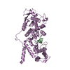
|
|---|---|
| 1 | x 15
|
-Components
| #1: Protein | Mass: 46273.625 Da / Num. of mol.: 1 Source method: isolated from a genetically manipulated source Source: (gene. exp.) Morbillivirus sp. / Production host:  |
|---|---|
| #2: RNA chain | Mass: 1930.277 Da / Num. of mol.: 1 Source method: isolated from a genetically manipulated source Source: (gene. exp.) Morbillivirus sp. / Production host: synthetic construct (others) |
-Experimental details
-Experiment
| Experiment | Method: ELECTRON MICROSCOPY |
|---|---|
| EM experiment | Aggregation state: HELICAL ARRAY / 3D reconstruction method: helical reconstruction |
- Sample preparation
| Component |
| ||||||||||||||||||||||||
|---|---|---|---|---|---|---|---|---|---|---|---|---|---|---|---|---|---|---|---|---|---|---|---|---|---|
| Molecular weight | Experimental value: NO | ||||||||||||||||||||||||
| Source (natural) |
| ||||||||||||||||||||||||
| Source (recombinant) |
| ||||||||||||||||||||||||
| Buffer solution | pH: 7.4 | ||||||||||||||||||||||||
| Buffer component |
| ||||||||||||||||||||||||
| Specimen | Conc.: 0.8 mg/ml / Embedding applied: NO / Shadowing applied: NO / Staining applied: NO / Vitrification applied: YES | ||||||||||||||||||||||||
| Specimen support | Grid material: COPPER / Grid type: Quantifoil R1.2/1.3 | ||||||||||||||||||||||||
| Vitrification | Instrument: FEI VITROBOT MARK IV / Cryogen name: ETHANE-PROPANE / Humidity: 95 % / Chamber temperature: 4 K |
- Electron microscopy imaging
| Experimental equipment |  Model: Titan Krios / Image courtesy: FEI Company |
|---|---|
| Microscopy | Model: TFS KRIOS |
| Electron gun | Electron source:  FIELD EMISSION GUN / Accelerating voltage: 300 kV / Illumination mode: FLOOD BEAM FIELD EMISSION GUN / Accelerating voltage: 300 kV / Illumination mode: FLOOD BEAM |
| Electron lens | Mode: BRIGHT FIELD / Nominal magnification: 22500 X / Nominal defocus max: 3000 nm / Nominal defocus min: 500 nm / Cs: 2.7 mm |
| Specimen holder | Cryogen: NITROGEN / Specimen holder model: FEI TITAN KRIOS AUTOGRID HOLDER |
| Image recording | Electron dose: 55 e/Å2 / Film or detector model: GATAN K3 (6k x 4k) / Num. of real images: 3153 |
- Processing
| EM software |
| ||||||||||||||||||
|---|---|---|---|---|---|---|---|---|---|---|---|---|---|---|---|---|---|---|---|
| CTF correction | Type: PHASE FLIPPING AND AMPLITUDE CORRECTION | ||||||||||||||||||
| Helical symmerty | Angular rotation/subunit: 28.5 ° / Axial rise/subunit: 5 Å / Axial symmetry: C1 | ||||||||||||||||||
| 3D reconstruction | Resolution: 4 Å / Resolution method: FSC 0.143 CUT-OFF / Num. of particles: 31361 / Symmetry type: HELICAL |
