 Movie
Movie Controller


+ Open data

- Basic information
| Entry | Database: PDB / ID: 7oho | ||||||
|---|---|---|---|---|---|---|---|
| Title | Crystal structure of AP2 FCHO2 chimera | ||||||
 Components Components | (AP-2 complex subunit ...) x 4 | ||||||
 Keywords Keywords | ENDOCYTOSIS / clathrin-mediated endocytosis (CME) / protein recycling / plasma membrane | ||||||
| Function / homology |  Function and homology information Function and homology informationmembrane invagination / Gap junction degradation / Formation of annular gap junctions / Nef Mediated CD8 Down-regulation / LDL clearance / WNT5A-dependent internalization of FZD2, FZD5 and ROR2 / WNT5A-dependent internalization of FZD2, FZD5 and ROR2 / WNT5A-dependent internalization of FZD4 / LDL clearance / Retrograde neurotrophin signalling ...membrane invagination / Gap junction degradation / Formation of annular gap junctions / Nef Mediated CD8 Down-regulation / LDL clearance / WNT5A-dependent internalization of FZD2, FZD5 and ROR2 / WNT5A-dependent internalization of FZD2, FZD5 and ROR2 / WNT5A-dependent internalization of FZD4 / LDL clearance / Retrograde neurotrophin signalling / VLDLR internalisation and degradation / WNT5A-dependent internalization of FZD2, FZD5 and ROR2 / Retrograde neurotrophin signalling / Trafficking of GluR2-containing AMPA receptors / WNT5A-dependent internalization of FZD4 / WNT5A-dependent internalization of FZD4 / clathrin adaptor complex / extrinsic component of presynaptic endocytic zone membrane / MHC class II antigen presentation / Trafficking of GluR2-containing AMPA receptors / VLDLR internalisation and degradation / Recycling pathway of L1 / regulation of vesicle size / postsynaptic endocytic zone / postsynaptic neurotransmitter receptor internalization / AP-2 adaptor complex / Retrograde neurotrophin signalling / Recycling pathway of L1 / Cargo recognition for clathrin-mediated endocytosis / membrane coat / clathrin-coated endocytic vesicle / Cargo recognition for clathrin-mediated endocytosis / clathrin coat assembly / LDL clearance / Clathrin-mediated endocytosis / positive regulation of synaptic vesicle endocytosis / Clathrin-mediated endocytosis / clathrin-cargo adaptor activity / vesicle budding from membrane / MHC class II antigen presentation / clathrin-dependent endocytosis / signal sequence receptor activity / Nef Mediated CD4 Down-regulation / coronary vasculature development / positive regulation of protein localization to membrane / endolysosome membrane / neurotransmitter secretion / clathrin-coated vesicle / Neutrophil degranulation / ventricular septum development / aorta development / low-density lipoprotein particle receptor binding / phosphatidylserine binding / clathrin binding / Trafficking of GluR2-containing AMPA receptors / Recycling pathway of L1 / positive regulation of receptor internalization / EPH-ephrin mediated repulsion of cells / positive regulation of endocytosis / negative regulation of protein localization to plasma membrane / synaptic vesicle endocytosis / vesicle-mediated transport / phosphatidylinositol-4,5-bisphosphate binding / clathrin-coated pit / MHC class II antigen presentation / phosphatidylinositol binding / VLDLR internalisation and degradation / protein serine/threonine kinase binding / protein localization to plasma membrane / kidney development / clathrin-coated endocytic vesicle membrane / intracellular protein transport / receptor internalization / kinase binding / cytoplasmic side of plasma membrane / disordered domain specific binding / terminal bouton / synaptic vesicle / endocytic vesicle membrane / Cargo recognition for clathrin-mediated endocytosis / Clathrin-mediated endocytosis / presynapse / protein-containing complex assembly / cytoplasmic vesicle / Potential therapeutics for SARS / transmembrane transporter binding / postsynapse / protein domain specific binding / lipid binding / synapse / protein kinase binding / protein-containing complex binding / glutamatergic synapse / membrane / identical protein binding / plasma membrane / cytoplasm / cytosol Similarity search - Function | ||||||
| Biological species |   Homo sapiens (human) Homo sapiens (human) | ||||||
| Method |  X-RAY DIFFRACTION / SYNCHROTRON / MOLECULAR REPLACEMENT / Resolution: 2.88 Å X-RAY DIFFRACTION / SYNCHROTRON / MOLECULAR REPLACEMENT / Resolution: 2.88 Å | ||||||
 Authors Authors | Zaccai, N.R. / Kelly, B.T. / Evans, P.R. / Owen, D.J. | ||||||
| Funding support |  United Kingdom, 1items United Kingdom, 1items
| ||||||
 Citation Citation | Journal: Sci Adv / Year: 2022 Title: FCHO controls AP2's initiating role in endocytosis through a PtdIns(4,5)P-dependent switch. Authors: Nathan R Zaccai / Zuzana Kadlecova / Veronica Kane Dickson / Kseniya Korobchevskaya / Jan Kamenicky / Oleksiy Kovtun / Perunthottathu K Umasankar / Antoni G Wrobel / Jonathan G G Kaufman / ...Authors: Nathan R Zaccai / Zuzana Kadlecova / Veronica Kane Dickson / Kseniya Korobchevskaya / Jan Kamenicky / Oleksiy Kovtun / Perunthottathu K Umasankar / Antoni G Wrobel / Jonathan G G Kaufman / Sally R Gray / Kun Qu / Philip R Evans / Marco Fritzsche / Filip Sroubek / Stefan Höning / John A G Briggs / Bernard T Kelly / David J Owen / Linton M Traub /     Abstract: Clathrin-mediated endocytosis (CME) is the main mechanism by which mammalian cells control their cell surface proteome. Proper operation of the pivotal CME cargo adaptor AP2 requires membrane- ...Clathrin-mediated endocytosis (CME) is the main mechanism by which mammalian cells control their cell surface proteome. Proper operation of the pivotal CME cargo adaptor AP2 requires membrane-localized Fer/Cip4 homology domain-only proteins (FCHO). Here, live-cell enhanced total internal reflection fluorescence-structured illumination microscopy shows that FCHO marks sites of clathrin-coated pit (CCP) initiation, which mature into uniform-sized CCPs comprising a central patch of AP2 and clathrin corralled by an FCHO/Epidermal growth factor potential receptor substrate number 15 (Eps15) ring. We dissect the network of interactions between the FCHO interdomain linker and AP2, which concentrates, orients, tethers, and partially destabilizes closed AP2 at the plasma membrane. AP2's subsequent membrane deposition drives its opening, which triggers FCHO displacement through steric competition with phosphatidylinositol 4,5-bisphosphate, clathrin, cargo, and CME accessory factors. FCHO can now relocate toward a CCP's outer edge to engage and activate further AP2s to drive CCP growth/maturation. #1: Journal: Acta Crystallogr., Sect. D: Biol. Crystallogr. / Year: 2012 Title: Towards automated crystallographic structure refinement with phenix.refine. Authors: Afonine, P.V. | ||||||
| History |
|
- Structure visualization
| Structure viewer | Molecule: MolmilJmol/JSmol |
|---|
- Downloads & links
-Download
| PDBx/mmCIF format | 7oho.cif.gz | 366.8 KB | Display | PDBx/mmCIF format |
|---|---|---|---|---|
| PDB format | pdb7oho.ent.gz | Display | PDB format | |
| PDBx/mmJSON format | 7oho.json.gz | Tree view | PDBx/mmJSON format | |
| Others |  Other downloads Other downloads |
-Validation report
| Arichive directory | https://data.pdbj.org/pub/pdb/validation_reports/oh/7ohoftp://data.pdbj.org/pub/pdb/validation_reports/oh/7oho | HTTPS FTP |
|---|
-Related structure data
| Related structure data | 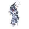 7ofpC 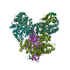 7og1C  7ohiC  7ohzC  7oi5C 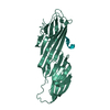 7oiqC  7oitC 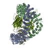 7z5cC C: citing same article ( |
|---|---|
| Similar structure data |


-Links
 PDBj
PDBj


- Assembly
| Deposited unit | 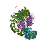
| ||||||||
|---|---|---|---|---|---|---|---|---|---|
| 1 |
| ||||||||
| Unit cell |
|
-Components
-AP-2 complex subunit ... , 4 types, 4 molecules AAABBBMMMSSS
| #1: Protein | Mass: 69656.297 Da / Num. of mol.: 1 Source method: isolated from a genetically manipulated source Source: (gene. exp.)  |
|---|---|
| #2: Protein | Mass: 71857.539 Da / Num. of mol.: 1 Source method: isolated from a genetically manipulated source Source: (gene. exp.) Homo sapiens (human) / Gene: AP2B1, ADTB2, CLAPB1, FCHO2 / Production host: |
| #3: Protein | Mass: 51044.113 Da / Num. of mol.: 1 Source method: isolated from a genetically manipulated source Source: (gene. exp.) |
| #4: Protein | Mass: 17038.688 Da / Num. of mol.: 1 Source method: isolated from a genetically manipulated source Source: (gene. exp.) |
-Non-polymers , 2 types, 5 molecules 

| #5: Chemical | ChemComp-IHP /  Mass: 660.035 Da / Num. of mol.: 1 / Source method: obtained synthetically / Formula: C6H18O24P6 Mass: 660.035 Da / Num. of mol.: 1 / Source method: obtained synthetically / Formula: C6H18O24P6 |
|---|---|
| #6: Chemical | ChemComp-GOL /  Mass: 92.094 Da / Num. of mol.: 4 / Source method: obtained synthetically / Formula: C3H8O3 Mass: 92.094 Da / Num. of mol.: 4 / Source method: obtained synthetically / Formula: C3H8O3 |
-Details
| Has ligand of interest | N |
|---|
-Experimental details
-Experiment
| Experiment | Method: X-RAY DIFFRACTION / Number of used crystals: 1 |
|---|
- Sample preparation
| Crystal | Density Matthews: 2.69 Å3/Da / Density % sol: 54.22 % |
|---|---|
| Crystal grow | Temperature: 289 K / Method: vapor diffusion, hanging drop / pH: 6.2 Details: 18% PEG 12000 0.1M Na/K phosphate pH 6.2 0.2M NaCl 4mM DTT in the presence of 3-fold molar excess of IP6. |
-Data collection
| Diffraction | Mean temperature: 100 K / Serial crystal experiment: N |
|---|---|
| Diffraction source | Source: SYNCHROTRON / Site: Diamond / Beamline: I02 / Wavelength: 0.9 Å |
| Detector | Type: DECTRIS PILATUS 6M-F / Detector: PIXEL / Date: Nov 9, 2015 |
| Radiation | Protocol: SINGLE WAVELENGTH / Monochromatic (M) / Laue (L): M / Scattering type: x-ray |
| Radiation wavelength | Wavelength: 0.9 Å / Relative weight: 1 |
| Reflection | Resolution: 2.88→66.61 Å / Num. obs: 51118 / % possible obs: 99.9 % / Redundancy: 9.8 % / Biso Wilson estimate: 79.944 Å2 / CC1/2: 0.998 / Rmerge(I) obs: 0.104 / Rpim(I) all: 0.037 / Rrim(I) all: 0.115 / Net I/σ(I): 13.5 |
| Reflection shell | Resolution: 2.88→2.95 Å / Redundancy: 9.6 % / Rmerge(I) obs: 1.803 / Mean I/σ(I) obs: 1.2 / Num. unique obs: 3710 / CC1/2: 0.529 / Rpim(I) all: 0.649 / Rrim(I) all: 2.019 / % possible all: 99.9 |
- Processing
| Software |
| |||||||||||||||||||||||||||||||||||||||||||||||||||||||||||||||||||||||||||||||||||||||||||||||||||||||||||||||||||||||||||||||||||||||||||||||||||||||||||
|---|---|---|---|---|---|---|---|---|---|---|---|---|---|---|---|---|---|---|---|---|---|---|---|---|---|---|---|---|---|---|---|---|---|---|---|---|---|---|---|---|---|---|---|---|---|---|---|---|---|---|---|---|---|---|---|---|---|---|---|---|---|---|---|---|---|---|---|---|---|---|---|---|---|---|---|---|---|---|---|---|---|---|---|---|---|---|---|---|---|---|---|---|---|---|---|---|---|---|---|---|---|---|---|---|---|---|---|---|---|---|---|---|---|---|---|---|---|---|---|---|---|---|---|---|---|---|---|---|---|---|---|---|---|---|---|---|---|---|---|---|---|---|---|---|---|---|---|---|---|---|---|---|---|---|---|---|
| Refinement | Method to determine structure: MOLECULAR REPLACEMENT Starting model: Phaser Resolution: 2.88→61.095 Å / Cor.coef. Fo:Fc: 0.964 / Cor.coef. Fo:Fc free: 0.925 / SU B: 20.665 / SU ML: 0.379 / Cross valid method: FREE R-VALUE / ESU R Free: 0.425 Details: Hydrogens have been added in their riding positions
| |||||||||||||||||||||||||||||||||||||||||||||||||||||||||||||||||||||||||||||||||||||||||||||||||||||||||||||||||||||||||||||||||||||||||||||||||||||||||||
| Solvent computation | Ion probe radii: 0.8 Å / Shrinkage radii: 0.8 Å / VDW probe radii: 1.2 Å / Solvent model: MASK BULK SOLVENT | |||||||||||||||||||||||||||||||||||||||||||||||||||||||||||||||||||||||||||||||||||||||||||||||||||||||||||||||||||||||||||||||||||||||||||||||||||||||||||
| Displacement parameters | Biso mean: 96.59 Å2
| |||||||||||||||||||||||||||||||||||||||||||||||||||||||||||||||||||||||||||||||||||||||||||||||||||||||||||||||||||||||||||||||||||||||||||||||||||||||||||
| Refinement step | Cycle: LAST / Resolution: 2.88→61.095 Å
| |||||||||||||||||||||||||||||||||||||||||||||||||||||||||||||||||||||||||||||||||||||||||||||||||||||||||||||||||||||||||||||||||||||||||||||||||||||||||||
| Refine LS restraints |
| |||||||||||||||||||||||||||||||||||||||||||||||||||||||||||||||||||||||||||||||||||||||||||||||||||||||||||||||||||||||||||||||||||||||||||||||||||||||||||
| LS refinement shell |
|
