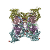
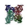
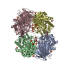
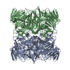
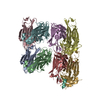
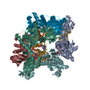
Journal: Elife / Year: 2021 Title: Structure and mechanistic features of the prokaryotic minimal RNase P. Authors: Rebecca Feyh / Nadine B Waeber / Simone Prinz / Pietro Ivan Giammarinaro / Gert Bange / Georg Hochberg / Roland K Hartmann / Florian Altegoer / Abstract: Endonucleolytic removal of 5'-leader sequences from tRNA precursor transcripts (pre-tRNAs) by ribonuclease P (RNase P) is essential for protein synthesis. Beyond RNA-based RNase P enzymes, protein- ...Endonucleolytic removal of 5'-leader sequences from tRNA precursor transcripts (pre-tRNAs) by ribonuclease P (RNase P) is essential for protein synthesis. Beyond RNA-based RNase P enzymes, protein-only versions of the enzyme exert this function in various eukarya (there termed PRORPs) and in some bacteria ( and close relatives); both enzyme types belong to distinct subgroups of the PIN domain metallonuclease superfamily. Homologs of RNase P (HARPs) are also expressed in some other bacteria and many archaea, where they coexist with RNA-based RNase P and do not represent the main RNase P activity. Here, we solved the structure of the bacterial HARP from by cryo-electron microscopy, revealing a novel screw-like dodecameric assembly. Biochemical experiments demonstrate that oligomerization is required for RNase P activity of HARPs. We propose that the tRNA substrate binds to an extended spike-helix (SH) domain that protrudes from the screw-like assembly to position the 5'-end in close proximity to the active site of the neighboring dimer. The structure suggests that eukaryotic PRORPs and prokaryotic HARPs recognize the same structural elements of pre-tRNAs (tRNA elbow region and cleavage site). Our analysis thus delivers the structural and mechanistic basis for pre-tRNA processing by the prokaryotic HARP system.
H: RNA-free ribonuclease P J: RNA-free ribonuclease P F: RNA-free ribonuclease P B: RNA-free ribonuclease P D: RNA-free ribonuclease P L: RNA-free ribonuclease P E: RNA-free ribonuclease P C: RNA-free ribonuclease P G: RNA-free ribonuclease P K: RNA-free ribonuclease P I: RNA-free ribonuclease P A: RNA-free ribonuclease P
Evidence: assay for oligomerization, Mass photometry was performed to determine the dynamic oligomeric assembly
Type
Name
Symmetry operation
Number
identity operation
1_555
1
Buried area
34590 Å2
ΔGint
-170 kcal/mol
Surface area
73650 Å2
Method
PISA
-
Components
#1: Protein
RNA-freeribonucleaseP / RNA-free RNase P / Protein-only RNase P
Mass: 24051.338 Da / Num. of mol.: 12 Source method: isolated from a genetically manipulated source Source: (gene. exp.) Halorhodospira halophila (strain DSM 244 / SL1) (bacteria) Strain: DSM 244 / SL1 / Gene: Hhal_2243 / Production host: Escherichia coli BL21(DE3) (bacteria) / References: UniProt: A1WZ95, ribonuclease P
-
Experimental details
-
Experiment
Experiment
Method: ELECTRON MICROSCOPY
EM experiment
Aggregation state: PARTICLE / 3D reconstruction method: single particle reconstruction
-
Sample preparation
Component
Name: Dodecameric assembly of minimal RNAseP system from Halorhodospira halophila Type: COMPLEX / Entity ID: all / Source: RECOMBINANT
Molecular weight
Value: 0.39 MDa / Experimental value: YES
Source (natural)
Organism: Halorhodospira halophila SL1 (bacteria)
Source (recombinant)
Organism: Escherichia coli BL21(DE3) (bacteria)
Buffer solution
pH: 8 Details: Solutions were prepared freshly and filtered through a 0.2 um filter
Buffer component
ID
Conc.
Name
Formula
Buffer-ID
1
100mM
potassiumchloride
KCl
1
2
20mM
Tris(hydroxymethyl)aminomethan
Tris
1
Specimen
Conc.: 8 mg/ml / Embedding applied: NO / Shadowing applied: NO / Staining applied: NO / Vitrification applied: YES / Details: The sample was monodisperse
Instrument: FEI VITROBOT MARK IV / Cryogen name: ETHANE / Humidity: 100 % / Chamber temperature: 283 K / Details: Blot for 11s with blot force -1 before plunging
-
Electron microscopy imaging
Experimental equipment
Model: Titan Krios / Image courtesy: FEI Company
Microscopy
Model: FEI TITAN KRIOS
Electron gun
Electron source: FIELD EMISSION GUN / Accelerating voltage: 300 kV / Illumination mode: FLOOD BEAM
In the structure databanks used in Yorodumi, some data are registered as the other names, "COVID-19 virus" and "2019-nCoV". Here are the details of the virus and the list of structure data.
Jan 31, 2019. EMDB accession codes are about to change! (news from PDBe EMDB page)
EMDB accession codes are about to change! (news from PDBe EMDB page)
The allocation of 4 digits for EMDB accession codes will soon come to an end. Whilst these codes will remain in use, new EMDB accession codes will include an additional digit and will expand incrementally as the available range of codes is exhausted. The current 4-digit format prefixed with “EMD-” (i.e. EMD-XXXX) will advance to a 5-digit format (i.e. EMD-XXXXX), and so on. It is currently estimated that the 4-digit codes will be depleted around Spring 2019, at which point the 5-digit format will come into force.
The EM Navigator/Yorodumi systems omit the EMD- prefix.
Related info.:Q: What is EMD? / ID/Accession-code notation in Yorodumi/EM Navigator
Yorodumi is a browser for structure data from EMDB, PDB, SASBDB, etc.
This page is also the successor to EM Navigator detail page, and also detail information page/front-end page for Omokage search.
The word "yorodu" (or yorozu) is an old Japanese word meaning "ten thousand". "mi" (miru) is to see.
Related info.:EMDB / PDB / SASBDB / Comparison of 3 databanks / Yorodumi Search / Aug 31, 2016. New EM Navigator & Yorodumi / Yorodumi Papers / Jmol/JSmol / Function and homology information / Changes in new EM Navigator and Yorodumi
 Movie
Movie Controller


 Open data
 Basic information
 Components
 Keywords
 Function and homology information
 Halorhodospira halophila (bacteria)
 Authors
 Citation
 Structure visualization
 Downloads & links
 Other downloads
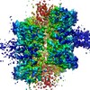

 PDBj
PDBj


 Assembly
 Sample preparation
 Electron microscopy imaging
 FIELD EMISSION GUN / Accelerating voltage: 300 kV / Illumination mode: FLOOD BEAM
 Processing