 Movie
Movie Controller

[English] 日本語
 Yorodumi
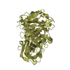
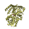
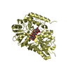
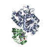
- PDB-7ofy: Crystal structure of SQ binding protein from Agrobacterium tumefa... -
+ Open data

- Basic information
| Entry | Database: PDB / ID: 7ofy | ||||||
|---|---|---|---|---|---|---|---|
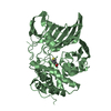
| Title | Crystal structure of SQ binding protein from Agrobacterium tumefaciens in complex with sulfoquinovosyl glycerol (SQGro) | ||||||
 Components Components | Sulfoquinovosyl binding protein | ||||||
 Keywords Keywords | SUGAR BINDING PROTEIN / sulfoquinovosyl diglyceride / SQDG / sulfoquinovose glycerol / SQGro / sulfoglycolysis | ||||||
| Function / homology | Bacterial extracellular solute-binding protein / Bacterial extracellular solute-binding protein / periplasmic space / Chem-VCW / Maltose-binding periplasmic protein Function and homology information Function and homology information | ||||||
| Biological species |  Rhizobium radiobacter (Agrobacterium genomosp. 4) Rhizobium radiobacter (Agrobacterium genomosp. 4) | ||||||
| Method |  X-RAY DIFFRACTION / SYNCHROTRON / MOLECULAR REPLACEMENT / molecular replacement / Resolution: 1.7 Å X-RAY DIFFRACTION / SYNCHROTRON / MOLECULAR REPLACEMENT / molecular replacement / Resolution: 1.7 Å | ||||||
 Authors Authors | Jarva, M.A. / Sharma, M. / Goddard-Borger, E.D. / Davies, G.J. | ||||||
| Funding support |  United Kingdom, 1items United Kingdom, 1items
| ||||||
 Citation Citation | Journal: Proc.Natl.Acad.Sci.USA / Year: 2022 Title: Oxidative desulfurization pathway for complete catabolism of sulfoquinovose by bacteria. Authors: Sharma, M. / Lingford, J.P. / Petricevic, M. / Snow, A.J.D. / Zhang, Y. / Jarva, M.A. / Mui, J.W. / Scott, N.E. / Saunders, E.C. / Mao, R. / Epa, R. / da Silva, B.M. / Pires, D.E.V. / ...Authors: Sharma, M. / Lingford, J.P. / Petricevic, M. / Snow, A.J.D. / Zhang, Y. / Jarva, M.A. / Mui, J.W. / Scott, N.E. / Saunders, E.C. / Mao, R. / Epa, R. / da Silva, B.M. / Pires, D.E.V. / Ascher, D.B. / McConville, M.J. / Davies, G.J. / Williams, S.J. / Goddard-Borger, E.D. | ||||||
| History |
|
- Structure visualization
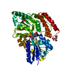
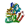
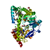
| Structure viewer | Molecule: MolmilJmol/JSmol |
|---|
- Downloads & links
-Download
| PDBx/mmCIF format | 7ofy.cif.gz | 321.7 KB | Display | PDBx/mmCIF format |
|---|---|---|---|---|
| PDB format | pdb7ofy.ent.gz | 260.4 KB | Display | PDB format |
| PDBx/mmJSON format | 7ofy.json.gz | Tree view | PDBx/mmJSON format | |
| Others |  Other downloads Other downloads |
-Validation report
| Arichive directory | https://data.pdbj.org/pub/pdb/validation_reports/of/7ofyftp://data.pdbj.org/pub/pdb/validation_reports/of/7ofy | HTTPS FTP |
|---|
-Related structure data
| Related structure data |  7bbyC 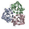 7bbzC 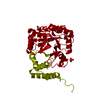 7bc0C  7bc1C  7nbzC 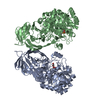 7ofxC  7oh2C  7olfC  6dtqS S: Starting model for refinement C: citing same article ( |
|---|---|
| Similar structure data |

-Links
 PDBj
PDBj


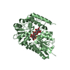
- Assembly
| Deposited unit | 
| ||||||||
|---|---|---|---|---|---|---|---|---|---|
| 1 | 
| ||||||||
| 2 | 
| ||||||||
| Unit cell |
|
-Components
| #1: Protein | Mass: 44074.840 Da / Num. of mol.: 2 Source method: isolated from a genetically manipulated source Source: (gene. exp.) Rhizobium radiobacter (Agrobacterium genomosp. 4)Gene: SY94_3278 / Production host: #2: Chemical |   Mass: 318.298 Da / Num. of mol.: 2 / Source method: obtained synthetically / Formula: C9H18O10S / Feature type: SUBJECT OF INVESTIGATION Mass: 318.298 Da / Num. of mol.: 2 / Source method: obtained synthetically / Formula: C9H18O10S / Feature type: SUBJECT OF INVESTIGATION#3: Chemical | ChemComp-EDO /   Mass: 62.068 Da / Num. of mol.: 10 / Source method: obtained synthetically / Formula: C2H6O2 Mass: 62.068 Da / Num. of mol.: 10 / Source method: obtained synthetically / Formula: C2H6O2#4: Water | ChemComp-HOH / |  Mass: 18.015 Da / Num. of mol.: 572 / Source method: isolated from a natural source / Formula: H2O Mass: 18.015 Da / Num. of mol.: 572 / Source method: isolated from a natural source / Formula: H2OHas ligand of interest | Y | |
|---|
-Experimental details
-Experiment
| Experiment | Method: X-RAY DIFFRACTION / Number of used crystals: 1 |
|---|
- Sample preparation
| Crystal | Density Matthews: 1.99 Å3/Da / Density % sol: 38.31 % |
|---|---|
| Crystal grow | Temperature: 293 K / Method: vapor diffusion Details: 32% PEG 4000, 0.2 M sodium acetate, 0.1 M Tris chloride, pH9 |
-Data collection
| Diffraction | Mean temperature: 100 K / Serial crystal experiment: N | |||||||||||||||||||||||||||
|---|---|---|---|---|---|---|---|---|---|---|---|---|---|---|---|---|---|---|---|---|---|---|---|---|---|---|---|---|
| Diffraction source | Source: SYNCHROTRON / Site: Australian Synchrotron  / Beamline: MX2 / Wavelength: 0.953664 Å / Beamline: MX2 / Wavelength: 0.953664 Å | |||||||||||||||||||||||||||
| Detector | Type: DECTRIS EIGER X 16M / Detector: PIXEL / Date: Oct 17, 2018 | |||||||||||||||||||||||||||
| Radiation | Protocol: SINGLE WAVELENGTH / Monochromatic (M) / Laue (L): M / Scattering type: x-ray | |||||||||||||||||||||||||||
| Radiation wavelength | Wavelength: 0.953664 Å / Relative weight: 1 | |||||||||||||||||||||||||||
| Reflection | Resolution: 1.7→47.19 Å / Num. obs: 74322 / % possible obs: 98.3 % / Redundancy: 3.4 % / Biso Wilson estimate: 19.39 Å2 / CC1/2: 0.998 / Rmerge(I) obs: 0.075 / Rpim(I) all: 0.048 / Rrim(I) all: 0.09 / Net I/σ(I): 9.8 / Num. measured all: 254947 / Scaling rejects: 1 | |||||||||||||||||||||||||||
| Reflection shell | Diffraction-ID: 1 / Redundancy: 3.4 %
|
-Phasing
| Phasing | Method: molecular replacement | |||||||||
|---|---|---|---|---|---|---|---|---|---|---|
| Phasing MR |
|
- Processing
| Software |
| ||||||||||||||||||||||||||||||||||||||||||||||||||||||||||||||||||||||||||||||||||||||||||||||||||||||||||||||||||||||||||||||||||||||||||||||||||||||||||||||||||
|---|---|---|---|---|---|---|---|---|---|---|---|---|---|---|---|---|---|---|---|---|---|---|---|---|---|---|---|---|---|---|---|---|---|---|---|---|---|---|---|---|---|---|---|---|---|---|---|---|---|---|---|---|---|---|---|---|---|---|---|---|---|---|---|---|---|---|---|---|---|---|---|---|---|---|---|---|---|---|---|---|---|---|---|---|---|---|---|---|---|---|---|---|---|---|---|---|---|---|---|---|---|---|---|---|---|---|---|---|---|---|---|---|---|---|---|---|---|---|---|---|---|---|---|---|---|---|---|---|---|---|---|---|---|---|---|---|---|---|---|---|---|---|---|---|---|---|---|---|---|---|---|---|---|---|---|---|---|---|---|---|---|---|---|
| Refinement | Method to determine structure: MOLECULAR REPLACEMENT Starting model: 6DTQ Resolution: 1.7→47.181 Å / SU ML: 0.19 / Cross valid method: THROUGHOUT / σ(F): 1.35 / Phase error: 18.87 / Stereochemistry target values: ML
| ||||||||||||||||||||||||||||||||||||||||||||||||||||||||||||||||||||||||||||||||||||||||||||||||||||||||||||||||||||||||||||||||||||||||||||||||||||||||||||||||||
| Solvent computation | Shrinkage radii: 0.9 Å / VDW probe radii: 1.11 Å / Solvent model: FLAT BULK SOLVENT MODEL | ||||||||||||||||||||||||||||||||||||||||||||||||||||||||||||||||||||||||||||||||||||||||||||||||||||||||||||||||||||||||||||||||||||||||||||||||||||||||||||||||||
| Displacement parameters | Biso max: 107.39 Å2 / Biso mean: 25.6471 Å2 / Biso min: 10.59 Å2 | ||||||||||||||||||||||||||||||||||||||||||||||||||||||||||||||||||||||||||||||||||||||||||||||||||||||||||||||||||||||||||||||||||||||||||||||||||||||||||||||||||
| Refinement step | Cycle: final / Resolution: 1.7→47.181 Å
| ||||||||||||||||||||||||||||||||||||||||||||||||||||||||||||||||||||||||||||||||||||||||||||||||||||||||||||||||||||||||||||||||||||||||||||||||||||||||||||||||||
| LS refinement shell | Refine-ID: X-RAY DIFFRACTION / Rfactor Rfree error: 0
| ||||||||||||||||||||||||||||||||||||||||||||||||||||||||||||||||||||||||||||||||||||||||||||||||||||||||||||||||||||||||||||||||||||||||||||||||||||||||||||||||||
| Refinement TLS params. | Method: refined / Refine-ID: X-RAY DIFFRACTION
| ||||||||||||||||||||||||||||||||||||||||||||||||||||||||||||||||||||||||||||||||||||||||||||||||||||||||||||||||||||||||||||||||||||||||||||||||||||||||||||||||||
| Refinement TLS group |
|
