 Movie
Movie Controller

[English] 日本語
 Yorodumi
- PDB-7bc1: Crystal structure of aldo-keto reductase from Agrobacterium tumef... -
+ Open data

- Basic information
| Entry | Database: PDB / ID: 7bc1 | ||||||
|---|---|---|---|---|---|---|---|
| Title | Crystal structure of aldo-keto reductase from Agrobacterium tumefaciens in a ternary complex with NADPH and glucose | ||||||
 Components Components | Aryl-alcohol dehydrogenase | ||||||
 Keywords Keywords | OXIDOREDUCTASE / Aldo-keto reductase / NADPH / glucose / sulfoquinovose | ||||||
| Function / homology |  Function and homology information Function and homology information | ||||||
| Biological species |  Agrobacterium fabrum (bacteria) Agrobacterium fabrum (bacteria) | ||||||
| Method |  X-RAY DIFFRACTION / SYNCHROTRON / MOLECULAR REPLACEMENT / Resolution: 1.5 Å X-RAY DIFFRACTION / SYNCHROTRON / MOLECULAR REPLACEMENT / Resolution: 1.5 Å | ||||||
 Authors Authors | Snow, A. / Sharma, M. / Davies, G.J. | ||||||
| Funding support |  United Kingdom, 1items United Kingdom, 1items
| ||||||
 Citation Citation | Journal: Proc.Natl.Acad.Sci.USA / Year: 2022 Title: Oxidative desulfurization pathway for complete catabolism of sulfoquinovose by bacteria. Authors: Sharma, M. / Lingford, J.P. / Petricevic, M. / Snow, A.J.D. / Zhang, Y. / Jarva, M.A. / Mui, J.W. / Scott, N.E. / Saunders, E.C. / Mao, R. / Epa, R. / da Silva, B.M. / Pires, D.E.V. / ...Authors: Sharma, M. / Lingford, J.P. / Petricevic, M. / Snow, A.J.D. / Zhang, Y. / Jarva, M.A. / Mui, J.W. / Scott, N.E. / Saunders, E.C. / Mao, R. / Epa, R. / da Silva, B.M. / Pires, D.E.V. / Ascher, D.B. / McConville, M.J. / Davies, G.J. / Williams, S.J. / Goddard-Borger, E.D. | ||||||
| History |
|
- Structure visualization
| Structure viewer | Molecule: MolmilJmol/JSmol |
|---|
- Downloads & links
-Download
| PDBx/mmCIF format | 7bc1.cif.gz | 78.5 KB | Display | PDBx/mmCIF format |
|---|---|---|---|---|
| PDB format | pdb7bc1.ent.gz | 54.6 KB | Display | PDB format |
| PDBx/mmJSON format | 7bc1.json.gz | Tree view | PDBx/mmJSON format | |
| Others |  Other downloads Other downloads |
-Validation report
| Arichive directory | https://data.pdbj.org/pub/pdb/validation_reports/bc/7bc1ftp://data.pdbj.org/pub/pdb/validation_reports/bc/7bc1 | HTTPS FTP |
|---|
-Related structure data
| Related structure data |  7bbySC 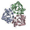 7bbzC 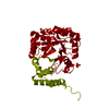 7bc0C  7nbzC 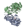 7ofxC  7ofyC  7oh2C  7olfC S: Starting model for refinement C: citing same article ( |
|---|---|
| Similar structure data |


-Links
 PDBj
PDBj


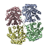
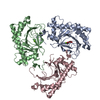
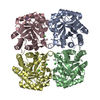
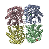
- Assembly
| Deposited unit | 
| ||||||||
|---|---|---|---|---|---|---|---|---|---|
| 1 | 
| ||||||||
| Unit cell |
|
-Components
| #1: Protein | Mass: 34326.129 Da / Num. of mol.: 1 Source method: isolated from a genetically manipulated source Source: (gene. exp.) Agrobacterium fabrum (strain C58 / ATCC 33970) (bacteria)Strain: C58 / ATCC 33970 / Gene: Atu3278 / Production host: |
|---|---|
| #2: Chemical | ChemComp-NAP /   Mass: 743.405 Da / Num. of mol.: 1 / Source method: obtained synthetically / Formula: C21H28N7O17P3 / Feature type: SUBJECT OF INVESTIGATION Mass: 743.405 Da / Num. of mol.: 1 / Source method: obtained synthetically / Formula: C21H28N7O17P3 / Feature type: SUBJECT OF INVESTIGATION |
| #3: Sugar | ChemComp-GLC /   Type: D-saccharide, alpha linking / Mass: 180.156 Da / Num. of mol.: 1 Type: D-saccharide, alpha linking / Mass: 180.156 Da / Num. of mol.: 1Source method: isolated from a genetically manipulated source Formula: C6H12O6 / Feature type: SUBJECT OF INVESTIGATION |
| #4: Water | ChemComp-HOH /  Mass: 18.015 Da / Num. of mol.: 229 / Source method: isolated from a natural source / Formula: H2O Mass: 18.015 Da / Num. of mol.: 229 / Source method: isolated from a natural source / Formula: H2O |
| Has ligand of interest | Y |
-Experimental details
-Experiment
| Experiment | Method: X-RAY DIFFRACTION / Number of used crystals: 1 |
|---|
- Sample preparation
| Crystal | Density Matthews: 2.2328458 Å3/Da / Density % sol: 44.947571 % / Description: Hexagonal plates |
|---|---|
| Crystal grow | Temperature: 293 K / Method: vapor diffusion, hanging drop / pH: 6 Details: 0.1 M SPG 25% PEG 1500 In-drop protein concentration of 13 mg/ml |
-Data collection
| Diffraction | Mean temperature: 100 K / Serial crystal experiment: N | ||||||||||||||||||||||||||||||
|---|---|---|---|---|---|---|---|---|---|---|---|---|---|---|---|---|---|---|---|---|---|---|---|---|---|---|---|---|---|---|---|
| Diffraction source | Source: SYNCHROTRON / Site: Diamond / Beamline: I04 / Wavelength: 0.9795 Å | ||||||||||||||||||||||||||||||
| Detector | Type: DECTRIS PILATUS 6M-F / Detector: PIXEL / Date: Apr 28, 2019 | ||||||||||||||||||||||||||||||
| Radiation | Protocol: SINGLE WAVELENGTH / Monochromatic (M) / Laue (L): M / Scattering type: x-ray | ||||||||||||||||||||||||||||||
| Radiation wavelength | Wavelength: 0.9795 Å / Relative weight: 1 | ||||||||||||||||||||||||||||||
| Reflection | Resolution: 1.5→52.57 Å / Num. obs: 48238 / % possible obs: 100 % / Redundancy: 11.9 % / CC1/2: 0.999 / Rmerge(I) obs: 0.082 / Rpim(I) all: 0.025 / Rrim(I) all: 0.086 / Net I/σ(I): 14.7 / Num. measured all: 571667 / Scaling rejects: 68 | ||||||||||||||||||||||||||||||
| Reflection shell | Diffraction-ID: 1
|
- Processing
| Software |
| ||||||||||||||||||||||||||||||||||||||||||||||||||||||||||||
|---|---|---|---|---|---|---|---|---|---|---|---|---|---|---|---|---|---|---|---|---|---|---|---|---|---|---|---|---|---|---|---|---|---|---|---|---|---|---|---|---|---|---|---|---|---|---|---|---|---|---|---|---|---|---|---|---|---|---|---|---|---|
| Refinement | Method to determine structure: MOLECULAR REPLACEMENT Starting model: 7BBY Resolution: 1.5→52.51 Å / Cor.coef. Fo:Fc: 0.977 / Cor.coef. Fo:Fc free: 0.969 / SU B: 2.001 / SU ML: 0.066 / Cross valid method: THROUGHOUT / σ(F): 0 / ESU R: 0.066 / ESU R Free: 0.069 / Stereochemistry target values: MAXIMUM LIKELIHOOD Details: HYDROGENS HAVE BEEN ADDED IN THE RIDING POSITIONS U VALUES : REFINED INDIVIDUALLY
| ||||||||||||||||||||||||||||||||||||||||||||||||||||||||||||
| Solvent computation | Ion probe radii: 0.8 Å / Shrinkage radii: 0.8 Å / VDW probe radii: 1.2 Å / Solvent model: MASK | ||||||||||||||||||||||||||||||||||||||||||||||||||||||||||||
| Displacement parameters | Biso max: 57.16 Å2 / Biso mean: 21.006 Å2 / Biso min: 13.7 Å2
| ||||||||||||||||||||||||||||||||||||||||||||||||||||||||||||
| Refinement step | Cycle: final / Resolution: 1.5→52.51 Å
| ||||||||||||||||||||||||||||||||||||||||||||||||||||||||||||
| Refine LS restraints |
| ||||||||||||||||||||||||||||||||||||||||||||||||||||||||||||
| LS refinement shell | Resolution: 1.5→1.539 Å / Rfactor Rfree error: 0 / Total num. of bins used: 20
|
