 Movie
Movie Controller

[English] 日本語
 Yorodumi
- PDB-7ofl: Crystal structure of the sesquiterpene synthase Copu9 from coniop... -
+ Open data

- Basic information
| Entry | Database: PDB / ID: 7ofl | ||||||
|---|---|---|---|---|---|---|---|
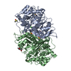
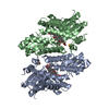
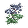
| Title | Crystal structure of the sesquiterpene synthase Copu9 from coniophora puteana in complex with alendronate | ||||||
 Components Components | Terpene synthase | ||||||
 Keywords Keywords | LYASE / terpene synthase / SESQUITERPENE / CYCLASE / TERPENOID | ||||||
| Function / homology |  Function and homology information Function and homology informationLyases; Carbon-oxygen lyases; Acting on phosphates / isoprenoid biosynthetic process / lyase activity / metal ion binding Similarity search - Function | ||||||
| Biological species | Coniophora puteana | ||||||
| Method |  X-RAY DIFFRACTION / SYNCHROTRON / MOLECULAR REPLACEMENT / Resolution: 1.83 Å X-RAY DIFFRACTION / SYNCHROTRON / MOLECULAR REPLACEMENT / Resolution: 1.83 Å | ||||||
 Authors Authors | Dimos, N. / Loll, B. | ||||||
 Citation Citation | Journal: Microb Cell Fact / Year: 2022 Title: Biotechnological potential and initial characterization of two novel sesquiterpene synthases from Basidiomycota Coniophora puteana for heterologous production of delta-cadinol. Authors: Ringel, M. / Dimos, N. / Himpich, S. / Haack, M. / Huber, C. / Eisenreich, W. / Schenk, G. / Loll, B. / Bruck, T. | ||||||
| History |
|
- Structure visualization
| Structure viewer | Molecule: MolmilJmol/JSmol |
|---|
- Downloads & links
-Download
| PDBx/mmCIF format | 7ofl.cif.gz | 204.8 KB | Display | PDBx/mmCIF format |
|---|---|---|---|---|
| PDB format | pdb7ofl.ent.gz | 129.9 KB | Display | PDB format |
| PDBx/mmJSON format | 7ofl.json.gz | Tree view | PDBx/mmJSON format | |
| Others |  Other downloads Other downloads |
-Validation report
| Arichive directory | https://data.pdbj.org/pub/pdb/validation_reports/of/7oflftp://data.pdbj.org/pub/pdb/validation_reports/of/7ofl | HTTPS FTP |
|---|
-Related structure data
| Related structure data | 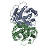 6tjzS S: Starting model for refinement |
|---|---|
| Similar structure data |


-Links
 PDBj
PDBj
- Assembly
| Deposited unit | 
| ||||||||||||
|---|---|---|---|---|---|---|---|---|---|---|---|---|---|
| 1 |
| ||||||||||||
| Unit cell |
| ||||||||||||
| Components on special symmetry positions |
|
-Components
| #1: Protein | Mass: 42807.289 Da / Num. of mol.: 2 Source method: isolated from a genetically manipulated source Source: (gene. exp.)  Coniophora puteana (strain RWD-64-598) (fungus) Coniophora puteana (strain RWD-64-598) (fungus)Strain: RWD-64-598 / Gene: CONPUDRAFT_118913 / Production host:  References: UniProt: A0A5M3MXY8, Lyases; Carbon-oxygen lyases; Acting on phosphates #2: Chemical |   Mass: 245.064 Da / Num. of mol.: 2 / Source method: obtained synthetically / Formula: C4H9NO7P2 Mass: 245.064 Da / Num. of mol.: 2 / Source method: obtained synthetically / Formula: C4H9NO7P2#3: Chemical | ChemComp-MG /   Mass: 24.305 Da / Num. of mol.: 6 / Source method: obtained synthetically / Formula: Mg Mass: 24.305 Da / Num. of mol.: 6 / Source method: obtained synthetically / Formula: Mg#4: Chemical | ChemComp-EDO /   Mass: 62.068 Da / Num. of mol.: 14 / Source method: obtained synthetically / Formula: C2H6O2 Mass: 62.068 Da / Num. of mol.: 14 / Source method: obtained synthetically / Formula: C2H6O2#5: Water | ChemComp-HOH / |  Mass: 18.015 Da / Num. of mol.: 697 / Source method: isolated from a natural source / Formula: H2O Mass: 18.015 Da / Num. of mol.: 697 / Source method: isolated from a natural source / Formula: H2OHas ligand of interest | N | |
|---|
-Experimental details
-Experiment
| Experiment | Method: X-RAY DIFFRACTION / Number of used crystals: 1 |
|---|
- Sample preparation
| Crystal | Density Matthews: 2.25 Å3/Da / Density % sol: 45.35 % |
|---|---|
| Crystal grow | Temperature: 277.15 K / Method: vapor diffusion, sitting drop / Details: 100mM Magnesium formate 15% PEG 3350 |
-Data collection
| Diffraction | Mean temperature: 100 K / Serial crystal experiment: N |
|---|---|
| Diffraction source | Source: SYNCHROTRON / Site: BESSY  / Beamline: 14.1 / Wavelength: 0.9184 Å / Beamline: 14.1 / Wavelength: 0.9184 Å |
| Detector | Type: DECTRIS PILATUS 6M / Detector: PIXEL / Date: Dec 12, 2020 |
| Radiation | Protocol: SINGLE WAVELENGTH / Monochromatic (M) / Laue (L): M / Scattering type: x-ray |
| Radiation wavelength | Wavelength: 0.9184 Å / Relative weight: 1 |
| Reflection | Resolution: 1.83→50 Å / Num. obs: 68244 / % possible obs: 98.6 % / Redundancy: 7.3 % / Biso Wilson estimate: 35.7 Å2 / CC1/2: 0.989 / Rrim(I) all: 0.525 / Net I/σ(I): 8.73 |
| Reflection shell | Resolution: 1.83→1.94 Å / Redundancy: 7.3 % / Mean I/σ(I) obs: 1.01 / Num. unique obs: 10301 / CC1/2: 0.44 / Rrim(I) all: 3.02 / % possible all: 93.3 |
- Processing
| Software |
| ||||||||||||||||||||||||||||||||||||||||||||||||||||||||||||||||||||||||||||||||||||||||||||||||||||||||||||||||
|---|---|---|---|---|---|---|---|---|---|---|---|---|---|---|---|---|---|---|---|---|---|---|---|---|---|---|---|---|---|---|---|---|---|---|---|---|---|---|---|---|---|---|---|---|---|---|---|---|---|---|---|---|---|---|---|---|---|---|---|---|---|---|---|---|---|---|---|---|---|---|---|---|---|---|---|---|---|---|---|---|---|---|---|---|---|---|---|---|---|---|---|---|---|---|---|---|---|---|---|---|---|---|---|---|---|---|---|---|---|---|---|---|---|
| Refinement | Method to determine structure: MOLECULAR REPLACEMENT Starting model: 6TJZ Resolution: 1.83→38.31 Å / SU ML: 0.2621 / Cross valid method: FREE R-VALUE / σ(F): 1.33 / Phase error: 25.185 Stereochemistry target values: GeoStd + Monomer Library + CDL v1.2
| ||||||||||||||||||||||||||||||||||||||||||||||||||||||||||||||||||||||||||||||||||||||||||||||||||||||||||||||||
| Solvent computation | Shrinkage radii: 0.9 Å / VDW probe radii: 1.11 Å / Solvent model: FLAT BULK SOLVENT MODEL | ||||||||||||||||||||||||||||||||||||||||||||||||||||||||||||||||||||||||||||||||||||||||||||||||||||||||||||||||
| Displacement parameters | Biso mean: 29.3 Å2 | ||||||||||||||||||||||||||||||||||||||||||||||||||||||||||||||||||||||||||||||||||||||||||||||||||||||||||||||||
| Refinement step | Cycle: LAST / Resolution: 1.83→38.31 Å
| ||||||||||||||||||||||||||||||||||||||||||||||||||||||||||||||||||||||||||||||||||||||||||||||||||||||||||||||||
| Refine LS restraints |
| ||||||||||||||||||||||||||||||||||||||||||||||||||||||||||||||||||||||||||||||||||||||||||||||||||||||||||||||||
| LS refinement shell |
|
