Movie
Movie Controller


+ Open data

- Basic information
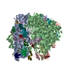
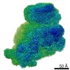
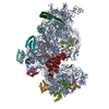
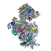
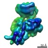
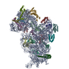
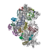
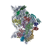
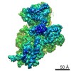
| Entry | Database: PDB / ID: 7oe1 | ||||||
|---|---|---|---|---|---|---|---|
| Title | 30S ribosomal subunit from E. coli | ||||||
 Components Components |
| ||||||
 Keywords Keywords | RIBOSOME / 30S subunit / RNA | ||||||
| Function / homology |  Function and homology information Function and homology informationmRNA 5'-UTR binding / small ribosomal subunit / small ribosomal subunit rRNA binding / cytosolic small ribosomal subunit / cytoplasmic translation / tRNA binding / rRNA binding / structural constituent of ribosome / ribosome / translation ...mRNA 5'-UTR binding / small ribosomal subunit / small ribosomal subunit rRNA binding / cytosolic small ribosomal subunit / cytoplasmic translation / tRNA binding / rRNA binding / structural constituent of ribosome / ribosome / translation / mRNA binding / cytoplasm / cytosol Similarity search - Function | ||||||
| Biological species |  | ||||||
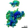
| Method | ELECTRON MICROSCOPY / single particle reconstruction / cryo EM / Resolution: 3.05 Å | ||||||
 Authors Authors | Maksimova, E. / Korepanov, A. / Baymukhametov, T. / Kravchenko, O. / Stolboushkina, E. | ||||||
| Funding support |  Russian Federation, 1items Russian Federation, 1items
| ||||||
 Citation Citation | Journal: Int J Mol Sci / Year: 2021 Title: RbfA Is Involved in Two Important Stages of 30S Subunit Assembly: Formation of the Central Pseudoknot and Docking of Helix 44 to the Decoding Center. Authors: Elena M Maksimova / Alexey P Korepanov / Olesya V Kravchenko / Timur N Baymukhametov / Alexander G Myasnikov / Konstantin S Vassilenko / Zhanna A Afonina / Elena A Stolboushkina /  Abstract: Ribosome biogenesis is a highly coordinated and complex process that requires numerous assembly factors that ensure prompt and flawless maturation of ribosomal subunits. Despite the increasing amount ...Ribosome biogenesis is a highly coordinated and complex process that requires numerous assembly factors that ensure prompt and flawless maturation of ribosomal subunits. Despite the increasing amount of data collected, the exact role of most assembly factors and mechanistic details of their operation remain unclear, mainly due to the shortage of high-resolution structural information. Here, using cryo-electron microscopy, we characterized 30S ribosomal particles isolated from an strain with a deleted gene for the RbfA factor. The cryo-EM maps for pre-30S subunits were divided into six classes corresponding to consecutive assembly intermediates: from the particles with a completely unresolved head domain and unfolded central pseudoknot to almost mature 30S subunits with well-resolved body, platform, and head domains and partially distorted helix 44. The structures of two predominant 30S intermediates belonging to most populated classes obtained at 2.7 Å resolutions indicate that RbfA acts at two distinctive 30S assembly stages: early formation of the central pseudoknot including folding of the head, and positioning of helix 44 in the decoding center at a later stage. Additionally, it was shown that the formation of the central pseudoknot may promote stabilization of the head domain, likely through the RbfA-dependent maturation of the neck helix 28. An update to the model of factor-dependent 30S maturation is proposed, suggesting that RfbA is involved in most of the subunit assembly process. | ||||||
| History |
|
- Structure visualization
| Movie |
Movie viewer |
|---|---|
| Structure viewer | Molecule: MolmilJmol/JSmol |
- Downloads & links
-Download
| PDBx/mmCIF format | 7oe1.cif.gz | 1.1 MB | Display | PDBx/mmCIF format |
|---|---|---|---|---|
| PDB format | pdb7oe1.ent.gz | 886.6 KB | Display | PDB format |
| PDBx/mmJSON format | 7oe1.json.gz | Tree view | PDBx/mmJSON format | |
| Others |  Other downloads Other downloads |
-Validation report
| Arichive directory | https://data.pdbj.org/pub/pdb/validation_reports/oe/7oe1ftp://data.pdbj.org/pub/pdb/validation_reports/oe/7oe1 | HTTPS FTP |
|---|
-Related structure data
| Related structure data | 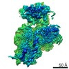 12857MC  7oe0C  7oi0C M: map data used to model this data C: citing same article ( |
|---|---|
| Similar structure data |


-Links
 PDBj
PDBj


- Assembly
| Deposited unit | 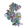
|
|---|---|
| 1 |
|
-Components
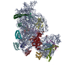
-RNA chain , 1 types, 1 molecules A
| #1: RNA chain | Mass: 499690.031 Da / Num. of mol.: 1 / Source method: isolated from a natural source Source: (natural) |
|---|
-30S ribosomal protein ... , 20 types, 20 molecules DEFHKLOPQRTBUCGIJMNS
| #2: Protein | Mass: 23383.002 Da / Num. of mol.: 1 / Source method: isolated from a natural source Source: (natural) References: UniProt: A0A6D2XM56 |
|---|---|
| #3: Protein | Mass: 17498.203 Da / Num. of mol.: 1 / Source method: isolated from a natural source Source: (natural) References: UniProt: A0A6D2YBZ5 |
| #4: Protein | Mass: 15727.512 Da / Num. of mol.: 1 / Source method: isolated from a natural source Source: (natural) References: UniProt: P02358 |
| #5: Protein | Mass: 14015.361 Da / Num. of mol.: 1 / Source method: isolated from a natural source Source: (natural) References: UniProt: A0A6D2XYQ3 |
| #6: Protein | Mass: 13739.778 Da / Num. of mol.: 1 Source method: isolated from a genetically manipulated source Source: (gene. exp.) Strain: K12 / Gene: rpsK, FAZ83_23205 Production host: References: UniProt: A0A6D2X4T2 |
| #7: Protein | Mass: 13636.961 Da / Num. of mol.: 1 / Source method: isolated from a natural source Source: (natural) References: UniProt: A0A4S5B3M5 |
| #8: Protein | Mass: 10319.882 Da / Num. of mol.: 1 / Source method: isolated from a natural source Source: (natural) References: UniProt: C3SSQ8 |
| #9: Protein | Mass: 9207.572 Da / Num. of mol.: 1 / Source method: isolated from a natural source Source: (natural) References: UniProt: A0A6D2XXS6 |
| #10: Protein | Mass: 9593.296 Da / Num. of mol.: 1 / Source method: isolated from a natural source Source: (natural) References: UniProt: A0A4S5APA8 |
| #11: Protein | Mass: 8874.276 Da / Num. of mol.: 1 / Source method: isolated from a natural source Source: (natural) References: UniProt: A0A6D2XHZ3 |
| #12: Protein | Mass: 9577.268 Da / Num. of mol.: 1 / Source method: isolated from a natural source Source: (natural) References: UniProt: A0A4S5B3X7 |
| #13: Protein | Mass: 26650.475 Da / Num. of mol.: 1 / Source method: isolated from a natural source Source: (natural) References: UniProt: A0A6D2W584 |
| #14: Protein | Mass: 8524.039 Da / Num. of mol.: 1 / Source method: isolated from a natural source Source: (natural) References: UniProt: A0A5F1E8X4 |
| #15: Protein | Mass: 25900.117 Da / Num. of mol.: 1 / Source method: isolated from a natural source Source: (natural) References: UniProt: A0A6D2XYP0 |
| #16: Protein | Mass: 19923.959 Da / Num. of mol.: 1 / Source method: isolated from a natural source Source: (natural) References: UniProt: C4ZUJ6 |
| #17: Protein | Mass: 14755.074 Da / Num. of mol.: 1 / Source method: isolated from a natural source Source: (natural) References: UniProt: A0A6D2XBM7 |
| #18: Protein | Mass: 11755.597 Da / Num. of mol.: 1 / Source method: isolated from a natural source Source: (natural) References: UniProt: A0A6D2XQX6 |
| #19: Protein | Mass: 12997.271 Da / Num. of mol.: 1 / Source method: isolated from a natural source Source: (natural) References: UniProt: A0A6D2XQ78 |
| #20: Protein | Mass: 11475.364 Da / Num. of mol.: 1 / Source method: isolated from a natural source Source: (natural) References: UniProt: A0A6D2WD65 |
| #21: Protein | Mass: 10324.160 Da / Num. of mol.: 1 / Source method: isolated from a natural source Source: (natural) References: UniProt: E3PL00 |
-Experimental details
-Experiment
| Experiment | Method: ELECTRON MICROSCOPY |
|---|---|
| EM experiment | Aggregation state: PARTICLE / 3D reconstruction method: single particle reconstruction |
- Sample preparation
| Component | Name: 30S ribosomal subunit from E.coli / Type: RIBOSOME / Entity ID: all / Source: NATURAL |
|---|---|
| Molecular weight | Value: 0.85 MDa |
| Source (natural) | Organism: |
| Buffer solution | pH: 7.5 |
| Specimen | Embedding applied: NO / Shadowing applied: NO / Staining applied: NO / Vitrification applied: YES |
| Vitrification | Cryogen name: ETHANE |
- Electron microscopy imaging
| Experimental equipment |  Model: Titan Krios / Image courtesy: FEI Company |
|---|---|
| Microscopy | Model: FEI TITAN KRIOS |
| Electron gun | Electron source:  FIELD EMISSION GUN / Accelerating voltage: 300 kV / Illumination mode: FLOOD BEAM FIELD EMISSION GUN / Accelerating voltage: 300 kV / Illumination mode: FLOOD BEAM |
| Electron lens | Mode: BRIGHT FIELD |
| Image recording | Electron dose: 2 e/Å2 / Film or detector model: FEI FALCON II (4k x 4k) |
- Processing
| EM software |
| ||||||||||||||||||||||||||||||||||||||||||||
|---|---|---|---|---|---|---|---|---|---|---|---|---|---|---|---|---|---|---|---|---|---|---|---|---|---|---|---|---|---|---|---|---|---|---|---|---|---|---|---|---|---|---|---|---|---|
| CTF correction | Type: PHASE FLIPPING ONLY | ||||||||||||||||||||||||||||||||||||||||||||
| 3D reconstruction | Resolution: 3.05 Å / Resolution method: FSC 0.143 CUT-OFF / Num. of particles: 169371 / Symmetry type: POINT | ||||||||||||||||||||||||||||||||||||||||||||
| Atomic model building | Protocol: RIGID BODY FIT / Space: REAL | ||||||||||||||||||||||||||||||||||||||||||||
| Atomic model building | 3D fitting-ID: 1 / Accession code: 4V4Q / Initial refinement model-ID: 1 / PDB-ID: 4V4Q / Source name: PDB / Type: experimental model
|