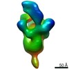
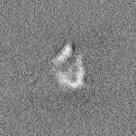
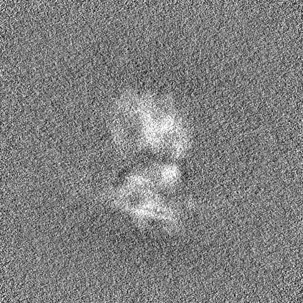
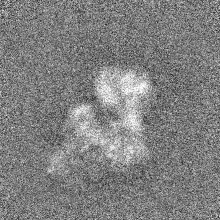
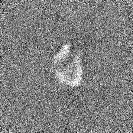
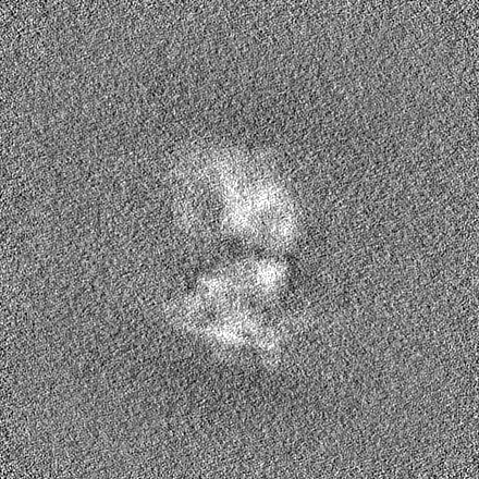
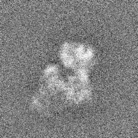
- EMDB-12855: E. coli pre-30S delta rbfA ribosomal subunit class A -
+
Open data
ID or keywords:
Loading...
-
Basic information
Entry
Database: EMDB / ID: EMD-12855
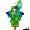
Title
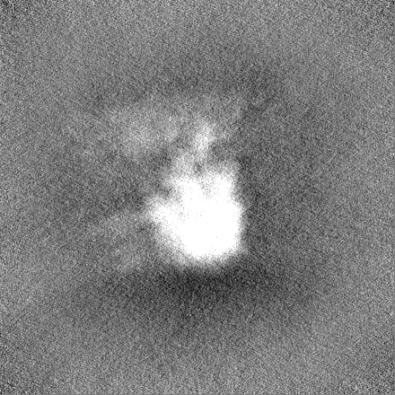
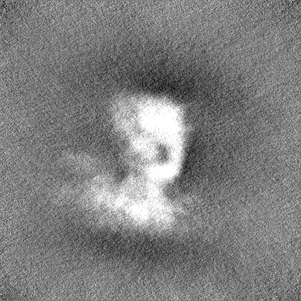
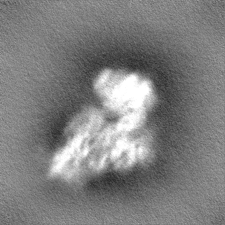
E. coli pre-30S delta rbfA ribosomal subunit class A
Map data
Sample
Complex: E.coli pre-30S delta rbfA ribosomal subunit class A
Function / homology
Function and homology information
mRNA 5'-UTR binding / small ribosomal subunit / small ribosomal subunit rRNA binding / cytosolic small ribosomal subunit / cytoplasmic translation / tRNA binding / rRNA binding / structural constituent of ribosome / ribosome / translation ...mRNA 5'-UTR binding / small ribosomal subunit / small ribosomal subunit rRNA binding / cytosolic small ribosomal subunit / cytoplasmic translation / tRNA binding / rRNA binding / structural constituent of ribosome / ribosome / translation / cytoplasm / cytosol Similarity search - Function
Ribosomal protein S16, conserved site / Ribosomal protein S16 signature. / Ribosomal protein S6, conserved site / Ribosomal protein S6 signature. / Ribosomal protein S11, bacterial-type / Ribosomal protein S20 / Ribosomal protein S20 superfamily / Ribosomal protein S20 / Ribosomal protein S4, bacterial-type / 30S ribosomal protein S17 ...Ribosomal protein S16, conserved site / Ribosomal protein S16 signature. / Ribosomal protein S6, conserved site / Ribosomal protein S6 signature. / Ribosomal protein S11, bacterial-type / Ribosomal protein S20 / Ribosomal protein S20 superfamily / Ribosomal protein S20 / Ribosomal protein S4, bacterial-type / 30S ribosomal protein S17 / Ribosomal protein S6, plastid/chloroplast / Ribosomal protein S18, conserved site / Ribosomal protein S18 signature. / Ribosomal protein S16 / Ribosomal protein S16 domain superfamily / Ribosomal protein S16 / Ribosomal protein S15, bacterial-type / Ribosomal protein S6 / Ribosomal protein S6 / Ribosomal protein S6 superfamily / Ribosomal protein S12, bacterial-type / Translation elongation factor EF1B/ribosomal protein S6 / Ribosomal protein S18 / Ribosomal protein S18 / Ribosomal protein S18 superfamily / Ribosomal protein S17, conserved site / Ribosomal protein S17 signature. / Ribosomal protein S4/S9 N-terminal domain / Ribosomal protein S8 signature. / Ribosomal protein S4, conserved site / Ribosomal protein S4 signature. / Ribosomal protein S4/S9 N-terminal domain / Ribosomal protein S4/S9, N-terminal / Ribosomal protein S15 signature. / Ribosomal protein S4/S9 / Ribosomal protein S8 / Ribosomal protein S8 superfamily / Ribosomal protein S8 / S4 RNA-binding domain profile. / S4 RNA-binding domain / S4 domain / Ribosomal S11, conserved site / Ribosomal protein S11 signature. / RNA-binding S4 domain / RNA-binding S4 domain superfamily / Ribosomal protein S11 / Ribosomal protein S12 signature. / Ribosomal protein S11 / Ribosomal protein S12/S23 / Ribosomal protein S12/S23 / Ribosomal protein S17/S11 / Ribosomal protein S17 / Ribosomal protein S15 / Ribosomal_S15 / Ribosomal protein S15 / Ribosomal protein S11 superfamily / S15/NS1, RNA-binding / Nucleic acid-binding, OB-fold Similarity search - Domain/homology
30S ribosomal protein S17 / 30S ribosomal protein S15 / 30S ribosomal protein S12 / 30S ribosomal protein S20 / 30S ribosomal protein S11 / 30S ribosomal protein S18 / 30S ribosomal protein S4 / 30S ribosomal protein S16 / 30S ribosomal protein S8 / Small ribosomal subunit protein bS6 Similarity search - Component
Biological species
Escherichia coli BW25113 (bacteria)
Method
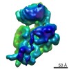
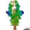
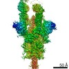
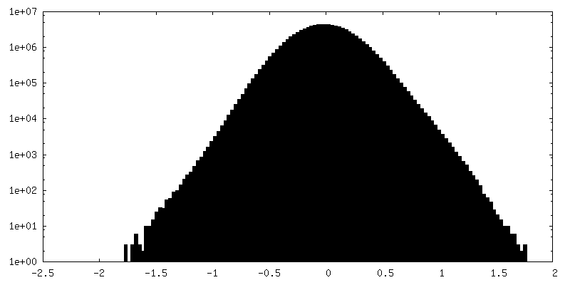
single particle reconstruction / cryo EM / Resolution: 12.0 Å
Journal: Int J Mol Sci / Year: 2021 Title: RbfA Is Involved in Two Important Stages of 30S Subunit Assembly: Formation of the Central Pseudoknot and Docking of Helix 44 to the Decoding Center. Authors: Elena M Maksimova / Alexey P Korepanov / Olesya V Kravchenko / Timur N Baymukhametov / Alexander G Myasnikov / Konstantin S Vassilenko / Zhanna A Afonina / Elena A Stolboushkina / Abstract: Ribosome biogenesis is a highly coordinated and complex process that requires numerous assembly factors that ensure prompt and flawless maturation of ribosomal subunits. Despite the increasing amount ...Ribosome biogenesis is a highly coordinated and complex process that requires numerous assembly factors that ensure prompt and flawless maturation of ribosomal subunits. Despite the increasing amount of data collected, the exact role of most assembly factors and mechanistic details of their operation remain unclear, mainly due to the shortage of high-resolution structural information. Here, using cryo-electron microscopy, we characterized 30S ribosomal particles isolated from an strain with a deleted gene for the RbfA factor. The cryo-EM maps for pre-30S subunits were divided into six classes corresponding to consecutive assembly intermediates: from the particles with a completely unresolved head domain and unfolded central pseudoknot to almost mature 30S subunits with well-resolved body, platform, and head domains and partially distorted helix 44. The structures of two predominant 30S intermediates belonging to most populated classes obtained at 2.7 Å resolutions indicate that RbfA acts at two distinctive 30S assembly stages: early formation of the central pseudoknot including folding of the head, and positioning of helix 44 in the decoding center at a later stage. Additionally, it was shown that the formation of the central pseudoknot may promote stabilization of the head domain, likely through the RbfA-dependent maturation of the neck helix 28. An update to the model of factor-dependent 30S maturation is proposed, suggesting that RfbA is involved in most of the subunit assembly process.
History
Deposition
Apr 30, 2021
-
Header (metadata) release
Jul 14, 2021
-
Map release
Jul 14, 2021
-
Update
Jul 14, 2021
-
Current status
Jul 14, 2021
Processing site: PDBe / Status: Released
-
Structure visualization
Movie
Surface view with section colored by density value
In the structure databanks used in Yorodumi, some data are registered as the other names, "COVID-19 virus" and "2019-nCoV". Here are the details of the virus and the list of structure data.
Jan 31, 2019. EMDB accession codes are about to change! (news from PDBe EMDB page)
EMDB accession codes are about to change! (news from PDBe EMDB page)
The allocation of 4 digits for EMDB accession codes will soon come to an end. Whilst these codes will remain in use, new EMDB accession codes will include an additional digit and will expand incrementally as the available range of codes is exhausted. The current 4-digit format prefixed with “EMD-” (i.e. EMD-XXXX) will advance to a 5-digit format (i.e. EMD-XXXXX), and so on. It is currently estimated that the 4-digit codes will be depleted around Spring 2019, at which point the 5-digit format will come into force.
The EM Navigator/Yorodumi systems omit the EMD- prefix.
Related info.:Q: What is EMD? / ID/Accession-code notation in Yorodumi/EM Navigator
Yorodumi is a browser for structure data from EMDB, PDB, SASBDB, etc.
This page is also the successor to EM Navigator detail page, and also detail information page/front-end page for Omokage search.
The word "yorodu" (or yorozu) is an old Japanese word meaning "ten thousand". "mi" (miru) is to see.
Related info.:EMDB / PDB / SASBDB / Comparison of 3 databanks / Yorodumi Search / Aug 31, 2016. New EM Navigator & Yorodumi / Yorodumi Papers / Jmol/JSmol / Function and homology information / Changes in new EM Navigator and Yorodumi
 Movie
Movie Controller


 Open data
 Basic information
 Map data
 Sample
 Function and homology information
 Authors
 Russian Federation, 1 items
  Citation
 Structure visualization
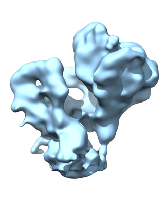
 Downloads & links
 emd_12855.png
 http://ftp.pdbj.org/pub/emdb/structures/EMD-12855


 Z (Sec.)
 Y (Row.)
 X (Col.)


 Sample components
 Processing
 Electron microscopy
 FIELD EMISSION GUN
