 Movie
Movie Controller


+ Open data

- Basic information
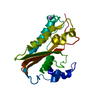
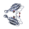
| Entry | Database: PDB / ID: 7ocz | ||||||
|---|---|---|---|---|---|---|---|
| Title | Crystal Structure of the PID-3 RRM domain | ||||||
 Components Components | Protein pid-3 | ||||||
 Keywords Keywords | RNA BINDING PROTEIN / piRNA biogenesis 21U RNA RNA recognition motif RNA binding | ||||||
| Function / homology |  Function and homology information Function and homology information21U-RNA metabolic process / positive regulation of chromosome segregation / RNA cap binding complex / piRNA processing / embryo development ending in birth or egg hatching / positive regulation of cell division / chromosome segregation / cell division / perinuclear region of cytoplasm / nucleus / cytoplasm Similarity search - Function | ||||||
| Biological species |  | ||||||
| Method |  X-RAY DIFFRACTION / SYNCHROTRON / MOLECULAR REPLACEMENT / Resolution: 1.82 Å X-RAY DIFFRACTION / SYNCHROTRON / MOLECULAR REPLACEMENT / Resolution: 1.82 Å | ||||||
 Authors Authors | Basquin, J. / Ketting, R.F. / Falk, S. | ||||||
 Citation Citation | Journal: Genes Dev. / Year: 2021 Title: Structural basis of PETISCO complex assembly during piRNA biogenesis in C. elegans . Authors: Perez-Borrajero, C. / Podvalnaya, N. / Holleis, K. / Lichtenberger, R. / Karaulanov, E. / Simon, B. / Basquin, J. / Hennig, J. / Ketting, R.F. / Falk, S. | ||||||
| History |
|
- Structure visualization
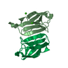
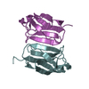
| Structure viewer | Molecule: MolmilJmol/JSmol |
|---|
- Downloads & links
-Download
| PDBx/mmCIF format | 7ocz.cif.gz | 55.1 KB | Display | PDBx/mmCIF format |
|---|---|---|---|---|
| PDB format | pdb7ocz.ent.gz | 31.8 KB | Display | PDB format |
| PDBx/mmJSON format | 7ocz.json.gz | Tree view | PDBx/mmJSON format | |
| Others |  Other downloads Other downloads |
-Validation report
| Arichive directory | https://data.pdbj.org/pub/pdb/validation_reports/oc/7oczftp://data.pdbj.org/pub/pdb/validation_reports/oc/7ocz | HTTPS FTP |
|---|
-Related structure data
| Related structure data | 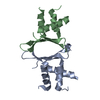 7o6lC 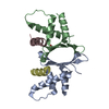 7o6nC  7ocxSC S: Starting model for refinement C: citing same article ( |
|---|---|
| Similar structure data |

-Links
 PDBj
PDBj- Assembly
| Deposited unit | 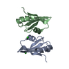
| ||||||||||||
|---|---|---|---|---|---|---|---|---|---|---|---|---|---|
| 1 |
| ||||||||||||
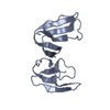
| Unit cell |
|
-Components
| #1: Protein | Mass: 9160.454 Da / Num. of mol.: 2 Source method: isolated from a genetically manipulated source Source: (gene. exp.)  #2: Chemical |   Mass: 35.453 Da / Num. of mol.: 2 / Source method: obtained synthetically / Formula: Cl Mass: 35.453 Da / Num. of mol.: 2 / Source method: obtained synthetically / Formula: Cl#3: Water | ChemComp-HOH / |  Mass: 18.015 Da / Num. of mol.: 106 / Source method: isolated from a natural source / Formula: H2O Mass: 18.015 Da / Num. of mol.: 106 / Source method: isolated from a natural source / Formula: H2OHas ligand of interest | N | |
|---|
-Experimental details
-Experiment
| Experiment | Method: X-RAY DIFFRACTION / Number of used crystals: 1 |
|---|
- Sample preparation
| Crystal | Density Matthews: 2.34 Å3/Da / Density % sol: 47.34 % |
|---|---|
| Crystal grow | Temperature: 295 K / Method: vapor diffusion, sitting drop Details: 0.03 M Sodium nitrate, 0.03 M Sodium phosphatedibasic 0.03 M Ammonium sulfate 0.1 M HEPES, 0.1 M MOPS pH 7.5 20% v/v PEG 500 MME; 10% w/v PEG 20000 |
-Data collection
| Diffraction | Mean temperature: 100 K / Serial crystal experiment: N |
|---|---|
| Diffraction source | Source: SYNCHROTRON / Site: SLS  / Beamline: X10SA / Wavelength: 1.28 Å / Beamline: X10SA / Wavelength: 1.28 Å |
| Detector | Type: DECTRIS EIGER X 16M / Detector: PIXEL / Date: Dec 11, 2020 |
| Radiation | Protocol: SINGLE WAVELENGTH / Monochromatic (M) / Laue (L): M / Scattering type: x-ray |
| Radiation wavelength | Wavelength: 1.28 Å / Relative weight: 1 |
| Reflection | Resolution: 1.82→37.49 Å / Num. obs: 14314 / % possible obs: 95.02 % / Redundancy: 17.9 % / Biso Wilson estimate: 24.56 Å2 / CC1/2: 0.987 / CC star: 0.997 / Net I/σ(I): 38.78 |
| Reflection shell | Resolution: 1.82→1.885 Å / Mean I/σ(I) obs: 0.56 / Num. unique obs: 1050 / CC1/2: 0.436 / CC star: 0.779 |
- Processing
| Software |
| |||||||||||||||||||||||||||||||||||||||||||||||||
|---|---|---|---|---|---|---|---|---|---|---|---|---|---|---|---|---|---|---|---|---|---|---|---|---|---|---|---|---|---|---|---|---|---|---|---|---|---|---|---|---|---|---|---|---|---|---|---|---|---|---|
| Refinement | Method to determine structure: MOLECULAR REPLACEMENT Starting model: 7OCX Resolution: 1.82→36.48 Å / SU ML: 0.2361 / Cross valid method: FREE R-VALUE / σ(F): 1.39 / Phase error: 28.6348 Stereochemistry target values: GeoStd + Monomer Library + CDL v1.2
| |||||||||||||||||||||||||||||||||||||||||||||||||
| Solvent computation | Shrinkage radii: 0.9 Å / VDW probe radii: 1.11 Å / Solvent model: FLAT BULK SOLVENT MODEL | |||||||||||||||||||||||||||||||||||||||||||||||||
| Displacement parameters | Biso mean: 31.77 Å2 | |||||||||||||||||||||||||||||||||||||||||||||||||
| Refinement step | Cycle: LAST / Resolution: 1.82→36.48 Å
| |||||||||||||||||||||||||||||||||||||||||||||||||
| Refine LS restraints |
| |||||||||||||||||||||||||||||||||||||||||||||||||
| LS refinement shell |
|
