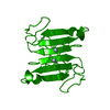
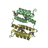
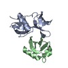
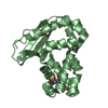
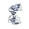
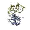
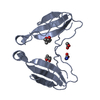
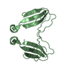
Entry Database : PDB / ID : 4aeaTitle Dimeric alpha-cobratoxin X-ray structure: Localization of intermolecular disulfides and possible mode of binding to nicotinic acetylcholine receptors LONG NEUROTOXIN 1 Keywords / / Function / homology Function Domain/homology Component
/ / / / / / / / / / / / Biological species NAJA KAOUTHIA (monocled cobra)Method / / / Resolution : 1.94 Å Authors Rucktooa, P. / Osipov, A.V. / Kasheverov, I.E. / Filkin, S.Y. / Starkov, V.G. / Andreeva, T.V. / Bertrand, D. / Utkin, Y.N. / Tsetlin, V.I. / Sixma, T.K. Journal : J.Biol.Chem. / Year : 2012Title : Dimeric Alpha-Cobratoxin X-Ray Structure: Localization of Intermolecular Disulfides and Possible Mode of Binding to Nicotinic Acetylcholine Receptors.Authors : Osipov, A.V. / Rucktooa, P. / Kasheverov, I.E. / Filkin, S.Y. / Starkov, V.G. / Andreeva, T.V. / Sixma, T.K. / Bertrand, D. / Utkin, Y.N. / Tsetlin, V.I. History Deposition Jan 9, 2012 Deposition site / Processing site Revision 1.0 Jan 25, 2012 Provider / Type Revision 1.1 Aug 15, 2012 Group Revision 1.2 Dec 20, 2023 Group Data collection / Database references ... Data collection / Database references / Derived calculations / Other / Refinement description Category chem_comp_atom / chem_comp_bond ... chem_comp_atom / chem_comp_bond / database_2 / pdbx_database_status / pdbx_initial_refinement_model / struct_site Item _database_2.pdbx_DOI / _database_2.pdbx_database_accession ... _database_2.pdbx_DOI / _database_2.pdbx_database_accession / _pdbx_database_status.status_code_sf / _struct_site.pdbx_auth_asym_id / _struct_site.pdbx_auth_comp_id / _struct_site.pdbx_auth_seq_id Revision 1.3 Nov 6, 2024 Group / Category / pdbx_modification_feature
Show all Show less
 Movie
Movie Controller

 Yorodumi
 Open data
 Basic information
 Components
 Keywords
 Function and homology information
 NAJA KAOUTHIA (monocled cobra)
 X-RAY DIFFRACTION /
  Authors
 Citation
 Structure visualization
 Downloads & links
 Other downloads
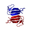


 PDBj
PDBj


 Assembly
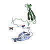

 Type: peptide linking / Mass: 75.067 Da / Num. of mol.: 1 / Source method: obtained synthetically / Formula: C2H5NO2
 Mass: 118.174 Da / Num. of mol.: 1 / Source method: obtained synthetically / Formula: C6H14O2 / Comment: precipitant*YM
 Mass: 18.015 Da / Num. of mol.: 69 / Source method: isolated from a natural source / Formula: H2O
 Sample preparation
 / Beamline: ID14-1 / Wavelength: 0.934
  Processing