 Movie
Movie Controller

[English] 日本語
 Yorodumi
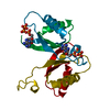
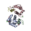
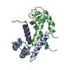
- PDB-1pfs: SOLUTION NMR STRUCTURE OF THE SINGLE-STRANDED DNA BINDING PROTEIN... -
+ Open data

- Basic information
| Entry | Database: PDB / ID: 1pfs | ||||||
|---|---|---|---|---|---|---|---|
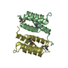
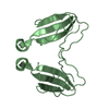
| Title | SOLUTION NMR STRUCTURE OF THE SINGLE-STRANDED DNA BINDING PROTEIN OF THE FILAMENTOUS PSEUDOMONAS PHAGE PF3, MINIMIZED AVERAGE STRUCTURE | ||||||
 Components Components | PF3 SINGLE-STRANDED DNA BINDING PROTEIN | ||||||
 Keywords Keywords | DNA BINDING PROTEIN / DNA-BINDING PROTEIN / VIRAL / BACTERIOPHAGE PF3 / SINGLE-STRANDED DNA | ||||||
| Function / homology |  Function and homology information Function and homology information | ||||||
| Biological species |  Pseudomonas phage Pf3 (virus) Pseudomonas phage Pf3 (virus) | ||||||
| Method | SOLUTION NMR | ||||||
 Authors Authors | Folmer, R.H.A. / Nilges, M. / Konings, R.N.H. / Hilbers, C.W. | ||||||
 Citation Citation | Journal: EMBO J. / Year: 1995 Title: Solution structure of the single-stranded DNA binding protein of the filamentous Pseudomonas phage Pf3: similarity to other proteins binding to single-stranded nucleic acids. Authors: Folmer, R.H. / Nilges, M. / Konings, R.N. / Hilbers, C.W. #1: Journal: Eur.J.Biochem. / Year: 1994Title: Secondary Structure of the Single-Stranded DNA Binding Protein Encoded by Filamentous Phage Pf3 as Determined by NMR Authors: Folmer, R.H. / Folkers, P.J. / Kaan, A. / Jonker, A.J. / Aelen, J.M. / Konings, R.N. / Hilbers, C.W. #2: Journal: J.Virol. / Year: 1985Title: Nucleotide Sequence of the Genome of Pf3, an Inc(P)-1 Plasmid-Specific Filamentous Bacteriophage of Pseudomonas Aeruginosa Authors: Luiten, R.G. / Putterman, D.G. / Schoenmakers, J.G. / Konings, R.N. / Day, L.A. | ||||||
| History |
|
- Structure visualization
| Structure viewer | Molecule: MolmilJmol/JSmol |
|---|
- Downloads & links
-Download
| PDBx/mmCIF format | 1pfs.cif.gz | 64 KB | Display | PDBx/mmCIF format |
|---|---|---|---|---|
| PDB format | pdb1pfs.ent.gz | 49.1 KB | Display | PDB format |
| PDBx/mmJSON format | 1pfs.json.gz | Tree view | PDBx/mmJSON format | |
| Others |  Other downloads Other downloads |
-Validation report
| Arichive directory | https://data.pdbj.org/pub/pdb/validation_reports/pf/1pfsftp://data.pdbj.org/pub/pdb/validation_reports/pf/1pfs | HTTPS FTP |
|---|
-Related structure data
| Similar structure data |
|---|

-Links
 PDBj
PDBj- Assembly
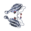
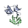
| Deposited unit | 
| |||||||||
|---|---|---|---|---|---|---|---|---|---|---|
| 1 |
| |||||||||
| NMR ensembles |
|
-Components
| #1: Protein | Mass: 8903.060 Da / Num. of mol.: 2 / Mutation: F36H Source method: isolated from a genetically manipulated source Details: DIMERIC / Source: (gene. exp.) Pseudomonas phage Pf3 (virus) / Genus: Inovirus / Plasmid: PPF3VF36H / Production host:  |
|---|
-Experimental details
-Experiment
| Experiment | Method: SOLUTION NMR | ||||||||||||||||||||||||||||||||
|---|---|---|---|---|---|---|---|---|---|---|---|---|---|---|---|---|---|---|---|---|---|---|---|---|---|---|---|---|---|---|---|---|---|
| NMR experiment |
|
- Sample preparation
| Sample conditions | pH: 4.4 / Temperature: 300 K |
|---|---|
| Crystal grow | *PLUS Method: other / Details: NMR |
-NMR measurement
| NMR spectrometer |
|
|---|
- Processing
| Software |
| ||||||||||||
|---|---|---|---|---|---|---|---|---|---|---|---|---|---|
| NMR software | Name:  X-PLOR / Version: 3.1 / Developer: BRUNGER / Classification: refinement X-PLOR / Version: 3.1 / Developer: BRUNGER / Classification: refinement | ||||||||||||
| Refinement | Software ordinal: 1 Details: 30 STRUCTURES WERE CALCULATED USING THE GEOMETRIC FORCE FIELD 'PARALLDHG.PRO'. THESE WERE SUBSEQUENTLY REFINED IN A 7 ANGSTROM SHELL OF WATER MOLECULES USING THE OPLS FORCE FIELD (TOPOPLSXX. ...Details: 30 STRUCTURES WERE CALCULATED USING THE GEOMETRIC FORCE FIELD 'PARALLDHG.PRO'. THESE WERE SUBSEQUENTLY REFINED IN A 7 ANGSTROM SHELL OF WATER MOLECULES USING THE OPLS FORCE FIELD (TOPOPLSXX.PRO) AND AVERAGED. THIS AVERAGED STRUCTURE WAS ENERGY MINIMIZED USING AGAIN 'PARALLDHG.PRO'. THE PROGRAM WAS MODIFIED TO USE FLOATING CHIRALITY. CHIRAL-CENTER RESTRAINT (A**3) : 0.352 | ||||||||||||
| NMR ensemble | Conformer selection criteria: OVERALL ENERGY / Conformers calculated total number: 80 / Conformers submitted total number: 1 |
