 Movie
Movie Controller

[English] 日本語
 Yorodumi
- PDB-7obk: Crystal structure of 14-3-3 sigma in complex with PKR phosphopeptide -
+ Open data

- Basic information
| Entry | Database: PDB / ID: 7obk | ||||||
|---|---|---|---|---|---|---|---|
| Title | Crystal structure of 14-3-3 sigma in complex with PKR phosphopeptide | ||||||
 Components Components |
| ||||||
 Keywords Keywords | SIGNALING PROTEIN / 14-3-3 protein protein-peptide complex | ||||||
| Function / homology |  Function and homology information Function and homology informationInhibition of PKR / regulation of NLRP3 inflammasome complex assembly / eukaryotic translation initiation factor 2alpha kinase activity / response to interferon-alpha / negative regulation of osteoblast proliferation / regulation of hematopoietic progenitor cell differentiation / positive regulation of stress-activated MAPK cascade / protein phosphatase regulator activity / SUMOylation of immune response proteins / regulation of hematopoietic stem cell proliferation ...Inhibition of PKR / regulation of NLRP3 inflammasome complex assembly / eukaryotic translation initiation factor 2alpha kinase activity / response to interferon-alpha / negative regulation of osteoblast proliferation / regulation of hematopoietic progenitor cell differentiation / positive regulation of stress-activated MAPK cascade / protein phosphatase regulator activity / SUMOylation of immune response proteins / regulation of hematopoietic stem cell proliferation / regulation of hematopoietic stem cell differentiation / regulation of translational initiation / negative regulation of viral genome replication / regulation of epidermal cell division / protein kinase C inhibitor activity / positive regulation of epidermal cell differentiation / keratinocyte development / keratinization / regulation of cell-cell adhesion / establishment of skin barrier / Regulation of localization of FOXO transcription factors / keratinocyte proliferation / negative regulation of keratinocyte proliferation / Activation of BAD and translocation to mitochondria / phosphoserine residue binding / cAMP/PKA signal transduction / negative regulation of protein localization to plasma membrane / SARS-CoV-2 targets host intracellular signalling and regulatory pathways / negative regulation of protein kinase activity / negative regulation of stem cell proliferation / SARS-CoV-1 targets host intracellular signalling and regulatory pathways / RHO GTPases activate PKNs / Chk1/Chk2(Cds1) mediated inactivation of Cyclin B:Cdk1 complex / positive regulation of protein localization / positive regulation of chemokine production / endoplasmic reticulum unfolded protein response / protein export from nucleus / TP53 Regulates Transcription of Genes Involved in G2 Cell Cycle Arrest / negative regulation of innate immune response / positive regulation of cell adhesion / release of cytochrome c from mitochondria / antiviral innate immune response / positive regulation of protein export from nucleus / stem cell proliferation / positive regulation of cytokine production / cellular response to amino acid starvation / : / TP53 Regulates Metabolic Genes / non-specific protein-tyrosine kinase / Translocation of SLC2A4 (GLUT4) to the plasma membrane / non-membrane spanning protein tyrosine kinase activity / protein sequestering activity / positive regulation of non-canonical NF-kappaB signal transduction / PKR-mediated signaling / Evasion by RSV of host interferon responses / ISG15 antiviral mechanism / intrinsic apoptotic signaling pathway in response to DNA damage / response to virus / kinase activity / Interferon alpha/beta signaling / intracellular protein localization / protein autophosphorylation / regulation of protein localization / double-stranded RNA binding / positive regulation of cell growth / sperm midpiece / defense response to virus / protein phosphorylation / protein kinase activity / positive regulation of MAPK cascade / non-specific serine/threonine protein kinase / regulation of cell cycle / negative regulation of translation / cadherin binding / ribosome / translation / negative regulation of cell population proliferation / protein serine kinase activity / protein serine/threonine kinase activity / protein kinase binding / negative regulation of apoptotic process / perinuclear region of cytoplasm / negative regulation of transcription by RNA polymerase II / signal transduction / : / RNA binding / extracellular exosome / nucleoplasm / ATP binding / membrane / identical protein binding / nucleus / cytoplasm / cytosol Similarity search - Function | ||||||
| Biological species |  Homo sapiens (human) Homo sapiens (human) | ||||||
| Method |  X-RAY DIFFRACTION / MOLECULAR REPLACEMENT / Resolution: 1.8 Å X-RAY DIFFRACTION / MOLECULAR REPLACEMENT / Resolution: 1.8 Å | ||||||
 Authors Authors | Centorrino, F. / Andlovic, B. / Ottmann, C. | ||||||
| Funding support | European Union, 1items
| ||||||
 Citation Citation | Journal: Cell Chem Biol / Year: 2023 Title: IFN alpha primes cancer cells for Fusicoccin-induced cell death via 14-3-3 PPI stabilization. Authors: Andlovic, B. / Heilmann, G. / Ninck, S. / Andrei, S.A. / Centorrino, F. / Higuchi, Y. / Kato, N. / Brunsveld, L. / Arkin, M. / Menninger, S. / Choidas, A. / Wolf, A. / Klebl, B. / Kaschani, ...Authors: Andlovic, B. / Heilmann, G. / Ninck, S. / Andrei, S.A. / Centorrino, F. / Higuchi, Y. / Kato, N. / Brunsveld, L. / Arkin, M. / Menninger, S. / Choidas, A. / Wolf, A. / Klebl, B. / Kaschani, F. / Kaiser, M. / Eickhoff, J. / Ottmann, C. | ||||||
| History |
|
- Structure visualization
| Structure viewer | Molecule: MolmilJmol/JSmol |
|---|
- Downloads & links
-Download
| PDBx/mmCIF format | 7obk.cif.gz | 142.5 KB | Display | PDBx/mmCIF format |
|---|---|---|---|---|
| PDB format | pdb7obk.ent.gz | 89.9 KB | Display | PDB format |
| PDBx/mmJSON format | 7obk.json.gz | Tree view | PDBx/mmJSON format | |
| Others |  Other downloads Other downloads |
-Validation report
| Arichive directory | https://data.pdbj.org/pub/pdb/validation_reports/ob/7obkftp://data.pdbj.org/pub/pdb/validation_reports/ob/7obk | HTTPS FTP |
|---|
-Related structure data
| Related structure data |  7ob5C  7ob8C  7obcC  7obdC  7obgC  7obhC 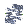 7oblC  7obsC 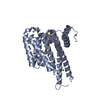 7obtC  7obxC 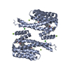 7obyC 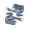 4jc3S S: Starting model for refinement C: citing same article ( |
|---|---|
| Similar structure data |
-Links
 PDBj
PDBj


- Assembly
| Deposited unit | 
| ||||||||||||
|---|---|---|---|---|---|---|---|---|---|---|---|---|---|
| 1 | 
| ||||||||||||
| Unit cell |
| ||||||||||||
| Components on special symmetry positions |
|
-Components
| #1: Protein | Mass: 28226.518 Da / Num. of mol.: 1 Source method: isolated from a genetically manipulated source Source: (gene. exp.) Homo sapiens (human) / Gene: SFN, HME1 / Production host:  | ||||||
|---|---|---|---|---|---|---|---|
| #2: Protein/peptide | Mass: 1428.465 Da / Num. of mol.: 1 / Source method: obtained synthetically / Source: (synth.) Homo sapiens (human)References: UniProt: P19525, non-specific serine/threonine protein kinase, non-specific protein-tyrosine kinase | ||||||
| #3: Chemical |   Mass: 24.305 Da / Num. of mol.: 2 / Source method: obtained synthetically / Formula: Mg Mass: 24.305 Da / Num. of mol.: 2 / Source method: obtained synthetically / Formula: Mg#4: Water | ChemComp-HOH / |  Mass: 18.015 Da / Num. of mol.: 442 / Source method: isolated from a natural source / Formula: H2O Mass: 18.015 Da / Num. of mol.: 442 / Source method: isolated from a natural source / Formula: H2OHas ligand of interest | Y | Has protein modification | Y | |
-Experimental details
-Experiment
| Experiment | Method: X-RAY DIFFRACTION / Number of used crystals: 1 |
|---|
- Sample preparation
| Crystal | Density Matthews: 2.46 Å3/Da / Density % sol: 49.95 % |
|---|---|
| Crystal grow | Temperature: 277.15 K / Method: vapor diffusion, sitting drop Details: 0.095 M Hepes pH7.3, 25%PEG 400, 0.19 M CaCl2 and 5 % Glycerol |
-Data collection
| Diffraction | Mean temperature: 100 K / Serial crystal experiment: N |
|---|---|
| Diffraction source | Source: SEALED TUBE / Type: RIGAKU MICROMAX-003 / Wavelength: 1.5419 Å |
| Detector | Type: DECTRIS PILATUS 200K / Detector: PIXEL / Date: Sep 1, 2020 |
| Radiation | Protocol: SINGLE WAVELENGTH / Monochromatic (M) / Laue (L): M / Scattering type: x-ray |
| Radiation wavelength | Wavelength: 1.5419 Å / Relative weight: 1 |
| Reflection | Resolution: 1.8→41.87 Å / Num. obs: 27306 / % possible obs: 99.8 % / Redundancy: 6.2 % / Biso Wilson estimate: 8.95 Å2 / CC1/2: 0.997 / Rmerge(I) obs: 0.088 / Net I/σ(I): 14.3 |
| Reflection shell | Resolution: 1.8→1.83 Å / Redundancy: 5 % / Rmerge(I) obs: 0.283 / Mean I/σ(I) obs: 4.5 / Num. unique obs: 1311 / CC1/2: 0.921 / % possible all: 97.8 |
- Processing
| Software |
| |||||||||||||||||||||||||||||||||||||||||||||||||||||||||||||||||||||||||||||
|---|---|---|---|---|---|---|---|---|---|---|---|---|---|---|---|---|---|---|---|---|---|---|---|---|---|---|---|---|---|---|---|---|---|---|---|---|---|---|---|---|---|---|---|---|---|---|---|---|---|---|---|---|---|---|---|---|---|---|---|---|---|---|---|---|---|---|---|---|---|---|---|---|---|---|---|---|---|---|
| Refinement | Method to determine structure: MOLECULAR REPLACEMENT Starting model: 4JC3 Resolution: 1.8→41.86 Å / SU ML: 0.1522 / Cross valid method: FREE R-VALUE / σ(F): 1.5 / Phase error: 16.1356 / Stereochemistry target values: GeoStd + Monomer Library
| |||||||||||||||||||||||||||||||||||||||||||||||||||||||||||||||||||||||||||||
| Solvent computation | Shrinkage radii: 0.9 Å / VDW probe radii: 1.11 Å / Solvent model: FLAT BULK SOLVENT MODEL | |||||||||||||||||||||||||||||||||||||||||||||||||||||||||||||||||||||||||||||
| Displacement parameters | Biso mean: 11.81 Å2 | |||||||||||||||||||||||||||||||||||||||||||||||||||||||||||||||||||||||||||||
| Refinement step | Cycle: LAST / Resolution: 1.8→41.86 Å
| |||||||||||||||||||||||||||||||||||||||||||||||||||||||||||||||||||||||||||||
| Refine LS restraints |
| |||||||||||||||||||||||||||||||||||||||||||||||||||||||||||||||||||||||||||||
| LS refinement shell |
|
