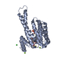
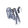
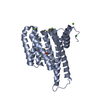
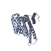
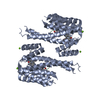
PDB-7ob5: Crystal structure of 14-3-3 sigma in complex with LDB1 phosphopeptide Method: X-RAY DIFFRACTION / Resolution: 1.8 Å
PDB-7ob8: Crystal structure of 14-3-3 sigma in complex with LDB1 phosphopeptide and stabilizer Fusicoccin-A Method: X-RAY DIFFRACTION / Resolution: 1.8 Å
PDB-7obc: Crystal structure of 14-3-3 sigma in complex with Phosphorylated and Farnesylated Rnd3 peptide Method: X-RAY DIFFRACTION / Resolution: 1.9 Å
PDB-7obd: Crystal structure of 14-3-3 sigma in complex with Phosphorylated and Farnesylated Rnd3 peptide and stabilizer Fusicoccin-A Method: X-RAY DIFFRACTION / Resolution: 2 Å
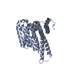
PDB-7obg: Crystal structure of 14-3-3 sigma in complex with NPM1 phosphopeptide Method: X-RAY DIFFRACTION / Resolution: 1.8 Å
PDB-7obh: Crystal structure of 14-3-3 sigma in complex with NPM1 phosphopeptide and stabilizer Fusicoccin-A Method: X-RAY DIFFRACTION / Resolution: 2 Å
PDB-7obk: Crystal structure of 14-3-3 sigma in complex with PKR phosphopeptide Method: X-RAY DIFFRACTION / Resolution: 1.8 Å
PDB-7obl: Crystal structure of 14-3-3 sigma in complex with PKR phosphopeptide and stabilizer Fusicoccin-A Method: X-RAY DIFFRACTION / Resolution: 1.8 Å
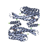
PDB-7obs: Crystal structure of 14-3-3 sigma in complex with RIPK2 phosphopeptide Method: X-RAY DIFFRACTION / Resolution: 1.8 Å
PDB-7obt: Crystal structure of 14-3-3 sigma in complex with RIPK2 phosphopeptide and stabilizer Fusicoccin-A Method: X-RAY DIFFRACTION / Resolution: 2.3 Å
PDB-7obx: Crystal structure of 14-3-3 sigma in complex with SSBP4 phosphopeptide Method: X-RAY DIFFRACTION / Resolution: 1.8 Å
PDB-7oby: Crystal structure of 14-3-3 sigma in complex with SSBP4 phosphopeptide and stabilizer Fusicoccin-A Method: X-RAY DIFFRACTION / Resolution: 2.1 Å
In the structure databanks used in Yorodumi, some data are registered as the other names, "COVID-19 virus" and "2019-nCoV". Here are the details of the virus and the list of structure data.
Jan 31, 2019. EMDB accession codes are about to change! (news from PDBe EMDB page)
EMDB accession codes are about to change! (news from PDBe EMDB page)
The allocation of 4 digits for EMDB accession codes will soon come to an end. Whilst these codes will remain in use, new EMDB accession codes will include an additional digit and will expand incrementally as the available range of codes is exhausted. The current 4-digit format prefixed with “EMD-” (i.e. EMD-XXXX) will advance to a 5-digit format (i.e. EMD-XXXXX), and so on. It is currently estimated that the 4-digit codes will be depleted around Spring 2019, at which point the 5-digit format will come into force.
The EM Navigator/Yorodumi systems omit the EMD- prefix.
Related info.:Q: What is EMD? / ID/Accession-code notation in Yorodumi/EM Navigator
 Movie
Movie Controller
 Structure viewers
 About Yorodumi Papers


 Authors
 External links


 Keywords
 homo sapiens (human)