 Movie
Movie Controller

[English] 日本語
 Yorodumi
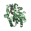
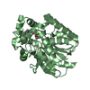
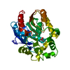
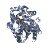
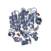
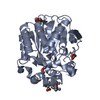
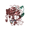
- PDB-7o8b: Structure of haloalkane dehalogenase variant DhaA80 from Rhodococ... -
+ Open data

- Basic information
| Entry | Database: PDB / ID: 7o8b | ||||||||||||||||||||||||
|---|---|---|---|---|---|---|---|---|---|---|---|---|---|---|---|---|---|---|---|---|---|---|---|---|---|
| Title | Structure of haloalkane dehalogenase variant DhaA80 from Rhodococcus rhodochrous | ||||||||||||||||||||||||
 Components Components | Haloalkane dehalogenase | ||||||||||||||||||||||||
 Keywords Keywords | HYDROLASE / alpha/beta hydrolase fold | ||||||||||||||||||||||||
| Function / homology |  Function and homology information Function and homology informationhaloalkane dehalogenase / haloalkane dehalogenase activity / response to toxic substance / membrane Similarity search - Function | ||||||||||||||||||||||||
| Biological species |  Rhodococcus rhodochrous (bacteria) Rhodococcus rhodochrous (bacteria) | ||||||||||||||||||||||||
| Method |  X-RAY DIFFRACTION / SYNCHROTRON / MOLECULAR REPLACEMENT / Resolution: 1.75 Å X-RAY DIFFRACTION / SYNCHROTRON / MOLECULAR REPLACEMENT / Resolution: 1.75 Å | ||||||||||||||||||||||||
 Authors Authors | Shaposhnikova, A. / Prudnikova, T. / Kuta Smatanova, I. | ||||||||||||||||||||||||
| Funding support |  Czech Republic, Czech Republic,  Germany, 7items Germany, 7items
| ||||||||||||||||||||||||
 Citation Citation | Journal: Crystals / Year: 2021 Title: Stabilization of Haloalkane Dehalogenase Structure by Interfacial Interaction with Ionic Liquids Authors: Shaposhnikova, A. / Kuty, M. / Chaloupkova, R. / Damborsky, J. / Kuta Smatanova, I. / Minofar, B. / Prudnikova, T. | ||||||||||||||||||||||||
| History |
|
- Structure visualization
| Structure viewer | Molecule: MolmilJmol/JSmol |
|---|
- Downloads & links
-Download
| PDBx/mmCIF format | 7o8b.cif.gz | 133.8 KB | Display | PDBx/mmCIF format |
|---|---|---|---|---|
| PDB format | pdb7o8b.ent.gz | 103.3 KB | Display | PDB format |
| PDBx/mmJSON format | 7o8b.json.gz | Tree view | PDBx/mmJSON format | |
| Others |  Other downloads Other downloads |
-Validation report
| Arichive directory | https://data.pdbj.org/pub/pdb/validation_reports/o8/7o8bftp://data.pdbj.org/pub/pdb/validation_reports/o8/7o8b | HTTPS FTP |
|---|
-Related structure data
| Related structure data |  7o3oC  4f60S S: Starting model for refinement C: citing same article ( |
|---|---|
| Similar structure data |
-Links
 PDBj
PDBj


- Assembly
| Deposited unit | 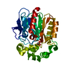
| ||||||||
|---|---|---|---|---|---|---|---|---|---|
| 1 |
| ||||||||
| Unit cell |
|
-Components
| #1: Protein | Mass: 33089.832 Da / Num. of mol.: 1 / Mutation: T148L, G171Q, A172V, C176F Source method: isolated from a genetically manipulated source Source: (gene. exp.) Rhodococcus rhodochrous (bacteria) / Gene: dhaA / Production host: |
|---|---|
| #2: Chemical | ChemComp-V5B / 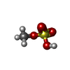  Mass: 112.105 Da / Num. of mol.: 1 / Source method: obtained synthetically / Formula: CH4O4S / Feature type: SUBJECT OF INVESTIGATION Mass: 112.105 Da / Num. of mol.: 1 / Source method: obtained synthetically / Formula: CH4O4S / Feature type: SUBJECT OF INVESTIGATION |
| #3: Water | ChemComp-HOH /  Mass: 18.015 Da / Num. of mol.: 133 / Source method: isolated from a natural source / Formula: H2O Mass: 18.015 Da / Num. of mol.: 133 / Source method: isolated from a natural source / Formula: H2O |
| Has ligand of interest | Y |
-Experimental details
-Experiment
| Experiment | Method: X-RAY DIFFRACTION / Number of used crystals: 1 |
|---|
- Sample preparation
| Crystal | Density Matthews: 2.18 Å3/Da / Density % sol: 43.66 % |
|---|---|
| Crystal grow | Temperature: 277 K / Method: vapor diffusion, sitting drop / pH: 6.5 Details: 0.1M BisTris propane, 0.2M Sodium Fluoride,20% PEG3350, 50% w/v 1-Butyl-3-methylimidazolium methyl sulfate |
-Data collection
| Diffraction | Mean temperature: 100 K / Serial crystal experiment: N |
|---|---|
| Diffraction source | Source: SYNCHROTRON / Site: BESSY / Beamline: 14.2 / Wavelength: 0.9184 Å |
| Detector | Type: MARMOSAIC 225 mm CCD / Detector: CCD / Date: Jan 29, 2015 |
| Radiation | Protocol: SINGLE WAVELENGTH / Monochromatic (M) / Laue (L): M / Scattering type: x-ray |
| Radiation wavelength | Wavelength: 0.9184 Å / Relative weight: 1 |
| Reflection | Resolution: 1.75→32.15 Å / Num. obs: 29090 / % possible obs: 99.17 % / Redundancy: 2 % / CC1/2: 0.989 / Net I/σ(I): 1.86 |
| Reflection shell | Resolution: 1.75→1.792 Å / Num. unique obs: 20 / Rrim(I) all: 0.369 |
- Processing
| Software |
| |||||||||||||||||||||||||||||||||||||||||||||||||||||||||||||||||||||||||||||||||||||||||||||||||||||||||
|---|---|---|---|---|---|---|---|---|---|---|---|---|---|---|---|---|---|---|---|---|---|---|---|---|---|---|---|---|---|---|---|---|---|---|---|---|---|---|---|---|---|---|---|---|---|---|---|---|---|---|---|---|---|---|---|---|---|---|---|---|---|---|---|---|---|---|---|---|---|---|---|---|---|---|---|---|---|---|---|---|---|---|---|---|---|---|---|---|---|---|---|---|---|---|---|---|---|---|---|---|---|---|---|---|---|---|
| Refinement | Method to determine structure: MOLECULAR REPLACEMENT Starting model: 4F60 Resolution: 1.75→32.15 Å / Cor.coef. Fo:Fc: 0.955 / Cor.coef. Fo:Fc free: 0.953 / SU B: 7.281 / SU ML: 0.107 / Cross valid method: THROUGHOUT / ESU R: 0.134 / ESU R Free: 0.117 / Stereochemistry target values: MAXIMUM LIKELIHOOD Details: U VALUES : WITH TLS ADDED HYDROGENS HAVE BEEN ADDED IN THE RIDING POSITIONS U VALUES : RESIDUAL ONLY
| |||||||||||||||||||||||||||||||||||||||||||||||||||||||||||||||||||||||||||||||||||||||||||||||||||||||||
| Solvent computation | Ion probe radii: 0.7 Å / Shrinkage radii: 0.7 Å / VDW probe radii: 1.2 Å / Solvent model: MASK | |||||||||||||||||||||||||||||||||||||||||||||||||||||||||||||||||||||||||||||||||||||||||||||||||||||||||
| Displacement parameters | Biso mean: 29.616 Å2
| |||||||||||||||||||||||||||||||||||||||||||||||||||||||||||||||||||||||||||||||||||||||||||||||||||||||||
| Refinement step | Cycle: LAST / Resolution: 1.75→32.15 Å
| |||||||||||||||||||||||||||||||||||||||||||||||||||||||||||||||||||||||||||||||||||||||||||||||||||||||||
| Refine LS restraints |
| |||||||||||||||||||||||||||||||||||||||||||||||||||||||||||||||||||||||||||||||||||||||||||||||||||||||||
| LS refinement shell | Resolution: 1.75→1.792 Å
| |||||||||||||||||||||||||||||||||||||||||||||||||||||||||||||||||||||||||||||||||||||||||||||||||||||||||
| Refinement TLS params. | Method: refined / Origin x: -10.156 Å / Origin y: 13.057 Å / Origin z: -17.473 Å
|
