 Movie
Movie Controller

[English] 日本語
 Yorodumi
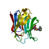
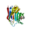
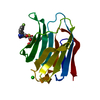
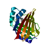
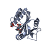
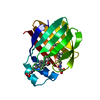
- PDB-7o60: Crystal structure of human myelin protein P2 at room temperature ... -
+ Open data

- Basic information
| Entry | Database: PDB / ID: 7o60 | ||||||
|---|---|---|---|---|---|---|---|
| Title | Crystal structure of human myelin protein P2 at room temperature from joint X-ray and neutron refinement. | ||||||
 Components Components | Myelin P2 protein | ||||||
 Keywords Keywords | LIPID BINDING PROTEIN / myelin / fatty acid binding protein / neuropathy / beta barrel | ||||||
| Function / homology |  Function and homology information Function and homology informationmembrane organization / cholesterol binding / fatty acid transport / fatty acid binding / myelin sheath / extracellular exosome / nucleus / cytosol Similarity search - Function | ||||||
| Biological species |  Homo sapiens (human) Homo sapiens (human) | ||||||
| Method |  X-RAY DIFFRACTION / NEUTRON DIFFRACTION / NUCLEAR REACTOR / MOLECULAR REPLACEMENT / Resolution: 2 Å X-RAY DIFFRACTION / NEUTRON DIFFRACTION / NUCLEAR REACTOR / MOLECULAR REPLACEMENT / Resolution: 2 Å | ||||||
 Authors Authors | Laulumaa, S. / Blakeley, M.P. / Kursula, P. | ||||||
 Citation Citation | Journal: Febs J. / Year: 2021 Title: Human myelin protein P2: from crystallography to time-lapse membrane imaging and neuropathy-associated variants. Authors: Uusitalo, M. / Klenow, M.B. / Laulumaa, S. / Blakeley, M.P. / Simonsen, A.C. / Ruskamo, S. / Kursula, P. | ||||||
| History |
|
- Structure visualization
| Structure viewer | Molecule: MolmilJmol/JSmol |
|---|
- Downloads & links
-Download
| PDBx/mmCIF format | 7o60.cif.gz | 93.2 KB | Display | PDBx/mmCIF format |
|---|---|---|---|---|
| PDB format | pdb7o60.ent.gz | 59.5 KB | Display | PDB format |
| PDBx/mmJSON format | 7o60.json.gz | Tree view | PDBx/mmJSON format | |
| Others |  Other downloads Other downloads |
-Validation report
| Arichive directory | https://data.pdbj.org/pub/pdb/validation_reports/o6/7o60ftp://data.pdbj.org/pub/pdb/validation_reports/o6/7o60 | HTTPS FTP |
|---|
-Related structure data
| Related structure data |  7nrwC  7nsrC  7ntpC  2wutS S: Starting model for refinement C: citing same article ( |
|---|---|
| Similar structure data |


-Links
 PDBj
PDBj


- Assembly
| Deposited unit | 
| ||||||||||||
|---|---|---|---|---|---|---|---|---|---|---|---|---|---|
| 1 |
| ||||||||||||
| Unit cell |
|
-Components
| #1: Protein | Mass: 14991.470 Da / Num. of mol.: 1 Source method: isolated from a genetically manipulated source Source: (gene. exp.) Homo sapiens (human) / Gene: PMP2 / Production host:  |
|---|---|
| #2: Chemical | ChemComp-CIT /   Mass: 192.124 Da / Num. of mol.: 1 / Source method: obtained synthetically / Formula: C6H8O7 Mass: 192.124 Da / Num. of mol.: 1 / Source method: obtained synthetically / Formula: C6H8O7 |
| #3: Chemical | ChemComp-PLM /   Mass: 256.424 Da / Num. of mol.: 1 / Source method: obtained synthetically / Formula: C16H32O2 Mass: 256.424 Da / Num. of mol.: 1 / Source method: obtained synthetically / Formula: C16H32O2 |
| #4: Chemical | ChemComp-DOD /   Mass: 18.015 Da / Num. of mol.: 105 / Source method: isolated from a natural source / Formula: D2O Mass: 18.015 Da / Num. of mol.: 105 / Source method: isolated from a natural source / Formula: D2O |
| Has ligand of interest | N |
| Has protein modification | Y |
-Experimental details
-Experiment
| Experiment |
|
|---|
- Sample preparation
| Crystal | Density Matthews: 2.82 Å3/Da / Density % sol: 56.42 % |
|---|---|
| Crystal grow | Temperature: 281 K / Method: vapor diffusion, hanging drop / Details: 28% PEG 6000, 0.1 M citrate, pD 4.75 |
-Data collection
| Diffraction |
| |||||||||||||||||||||||||||
|---|---|---|---|---|---|---|---|---|---|---|---|---|---|---|---|---|---|---|---|---|---|---|---|---|---|---|---|---|
| Diffraction source |
| |||||||||||||||||||||||||||
| Detector |
| |||||||||||||||||||||||||||
| Radiation |
| |||||||||||||||||||||||||||
| Radiation wavelength |
| |||||||||||||||||||||||||||
| Reflection | Biso Wilson estimate: 27.66 Å2 / Entry-ID: 7O60
| |||||||||||||||||||||||||||
| Reflection shell |
|

- Processing
| Software |
| ||||||||||||||||||||||||||||||||||||||||||||||||||||||||||||||||||||||
|---|---|---|---|---|---|---|---|---|---|---|---|---|---|---|---|---|---|---|---|---|---|---|---|---|---|---|---|---|---|---|---|---|---|---|---|---|---|---|---|---|---|---|---|---|---|---|---|---|---|---|---|---|---|---|---|---|---|---|---|---|---|---|---|---|---|---|---|---|---|---|---|
| Refinement | SU ML: 0.167 / Cross valid method: FREE R-VALUE / Method to determine structure:
| ||||||||||||||||||||||||||||||||||||||||||||||||||||||||||||||||||||||
| Refinement step | Cycle: LAST / Resolution: 2→14.53 Å
| ||||||||||||||||||||||||||||||||||||||||||||||||||||||||||||||||||||||
| Refine LS restraints |
| ||||||||||||||||||||||||||||||||||||||||||||||||||||||||||||||||||||||
| LS refinement shell |
|
