 Movie
Movie Controller

[English] 日本語
 Yorodumi
- PDB-7o5w: Crystal structure of holo-F210W mutant of Hydroxy ketone aldolase... -
+ Open data

- Basic information
| Entry | Database: PDB / ID: 7o5w | ||||||
|---|---|---|---|---|---|---|---|
| Title | Crystal structure of holo-F210W mutant of Hydroxy ketone aldolase (SwHKA)from Sphingomonas wittichii RW1 | ||||||
 Components Components | HpcH/HpaI aldolase | ||||||
 Keywords Keywords | LYASE / Class II pyruvate aldolase / Metal dependent aldolase / aldol reaction / Magnesium / carbon bond formation / holo / mutant | ||||||
| Function / homology | HpcH/HpaI aldolase/citrate lyase domain / HpcH/HpaI aldolase/citrate lyase / Pyruvate kinase-like domain superfamily / Pyruvate/Phosphoenolpyruvate kinase-like domain superfamily / catalytic activity / BROMIDE ION / : / DI(HYDROXYETHYL)ETHER / HpcH/HpaI aldolase Function and homology information Function and homology information | ||||||
| Biological species |  Sphingomonas wittichii (bacteria) Sphingomonas wittichii (bacteria) | ||||||
| Method |  X-RAY DIFFRACTION / SYNCHROTRON / MOLECULAR REPLACEMENT / Resolution: 1.2 Å X-RAY DIFFRACTION / SYNCHROTRON / MOLECULAR REPLACEMENT / Resolution: 1.2 Å | ||||||
 Authors Authors | Laustsen, J. / Justo, I. / Marsden, S.R. / Hanefeld, U. / Bento, I. | ||||||
 Citation Citation | Journal: Angew.Chem.Int.Ed.Engl. / Year: 2022 Title: Substrate Induced Movement of the Metal Cofactor between Active and Resting State. Authors: Marsden, S.R. / Wijma, H.J. / Mohr, M.K.F. / Justo, I. / Hagedoorn, P.L. / Laustsen, J. / Jeffries, C.M. / Svergun, D. / Mestrom, L. / McMillan, D.G.G. / Bento, I. / Hanefeld, U. | ||||||
| History |
|
- Structure visualization
| Structure viewer | Molecule: MolmilJmol/JSmol |
|---|
- Downloads & links
-Download
| PDBx/mmCIF format | 7o5w.cif.gz | 366.9 KB | Display | PDBx/mmCIF format |
|---|---|---|---|---|
| PDB format | pdb7o5w.ent.gz | Display | PDB format | |
| PDBx/mmJSON format | 7o5w.json.gz | Tree view | PDBx/mmJSON format | |
| Others |  Other downloads Other downloads |
-Validation report
| Arichive directory | https://data.pdbj.org/pub/pdb/validation_reports/o5/7o5wftp://data.pdbj.org/pub/pdb/validation_reports/o5/7o5w | HTTPS FTP |
|---|
-Related structure data
| Related structure data | 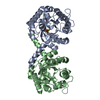 7nnkC 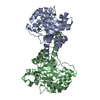 7nr1C 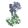 7nujC  7o5iC  7o5rC 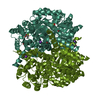 7o5vC 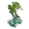 7o87C 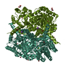 7o9rC 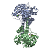 7obuC 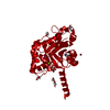 8adqC 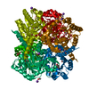 6r62S C: citing same article ( S: Starting model for refinement |
|---|---|
| Similar structure data |
-Links
 PDBj
PDBj


- Assembly
| Deposited unit | 
| |||||||||||||||||||||||||||||||||
|---|---|---|---|---|---|---|---|---|---|---|---|---|---|---|---|---|---|---|---|---|---|---|---|---|---|---|---|---|---|---|---|---|---|---|
| 1 | 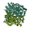
| |||||||||||||||||||||||||||||||||
| Unit cell |
| |||||||||||||||||||||||||||||||||
| Components on special symmetry positions |
| |||||||||||||||||||||||||||||||||
| Noncrystallographic symmetry (NCS) | NCS domain:
NCS domain segments: Component-ID: 1 / Ens-ID: 1 / Beg auth comp-ID: HIS / Beg label comp-ID: HIS / End auth comp-ID: ARG / End label comp-ID: ARG / Auth seq-ID: 1 - 249 / Label seq-ID: 2 - 250
NCS ensembles : (Details: Local NCS retraints between domains: 1 2) |
-Components
-Protein , 1 types, 2 molecules AAABBB
| #1: Protein | Mass: 26823.561 Da / Num. of mol.: 2 / Mutation: F210W Source method: isolated from a genetically manipulated source Source: (gene. exp.) Sphingomonas wittichii (strain RW1 / DSM 6014 / JCM 10273) (bacteria)Strain: RW1 / DSM 6014 / JCM 10273 / Gene: Swit_5035 / Production host: |
|---|
-Non-polymers , 5 types, 617 molecules 


| #2: Chemical | ChemComp-PEG /  Mass: 106.120 Da / Num. of mol.: 5 / Source method: obtained synthetically / Formula: C4H10O3 Mass: 106.120 Da / Num. of mol.: 5 / Source method: obtained synthetically / Formula: C4H10O3#3: Chemical |  Mass: 24.305 Da / Num. of mol.: 2 / Source method: obtained synthetically / Formula: Mg / Feature type: SUBJECT OF INVESTIGATION Mass: 24.305 Da / Num. of mol.: 2 / Source method: obtained synthetically / Formula: Mg / Feature type: SUBJECT OF INVESTIGATION#4: Chemical |  Mass: 79.904 Da / Num. of mol.: 3 / Source method: obtained synthetically / Formula: Br Mass: 79.904 Da / Num. of mol.: 3 / Source method: obtained synthetically / Formula: Br#5: Chemical |  Mass: 39.098 Da / Num. of mol.: 2 / Source method: obtained synthetically / Formula: K Mass: 39.098 Da / Num. of mol.: 2 / Source method: obtained synthetically / Formula: K#6: Water | ChemComp-HOH / | Mass: 18.015 Da / Num. of mol.: 605 / Source method: isolated from a natural source / Formula: H2O |
|---|
-Details
| Has ligand of interest | Y |
|---|
-Experimental details
-Experiment
| Experiment | Method: X-RAY DIFFRACTION / Number of used crystals: 1 |
|---|
- Sample preparation
| Crystal | Density Matthews: 1.91 Å3/Da / Density % sol: 35.76 % |
|---|---|
| Crystal grow | Temperature: 277.15 K / Method: vapor diffusion, hanging drop / pH: 7 / Details: HEPES, Sodium Citrate |
-Data collection
| Diffraction | Mean temperature: 100 K / Serial crystal experiment: N | |||||||||||||||||||||
|---|---|---|---|---|---|---|---|---|---|---|---|---|---|---|---|---|---|---|---|---|---|---|
| Diffraction source | Source: SYNCHROTRON / Site: PETRA III, EMBL c/o DESY  / Beamline: P13 (MX1) / Wavelength: 0.9762 Å / Beamline: P13 (MX1) / Wavelength: 0.9762 Å | |||||||||||||||||||||
| Detector | Type: DECTRIS PILATUS 6M-F / Detector: PIXEL / Date: Apr 26, 2019 | |||||||||||||||||||||
| Radiation | Monochromator: Oxforf-FNB / Protocol: SINGLE WAVELENGTH / Monochromatic (M) / Laue (L): M / Scattering type: x-ray | |||||||||||||||||||||
| Radiation wavelength | Wavelength: 0.9762 Å / Relative weight: 1 | |||||||||||||||||||||
| Reflection | Resolution: 1.2→74.27 Å / Num. obs: 130983 / % possible obs: 100 % / Redundancy: 9.7 % / CC1/2: 0.999 / Rmerge(I) obs: 0.078 / Rpim(I) all: 0.039 / Rrim(I) all: 0.087 / Net I/σ(I): 15.5 | |||||||||||||||||||||
| Reflection shell | Diffraction-ID: 1
|
- Processing
| Software |
| |||||||||||||||||||||||||||||||||||||||||||||||||||||||||||||||||||||||||||||||||||||||||||||||||||||||||||||||||||||||||||||||||||||||||||||||||||||||||||||||||||||
|---|---|---|---|---|---|---|---|---|---|---|---|---|---|---|---|---|---|---|---|---|---|---|---|---|---|---|---|---|---|---|---|---|---|---|---|---|---|---|---|---|---|---|---|---|---|---|---|---|---|---|---|---|---|---|---|---|---|---|---|---|---|---|---|---|---|---|---|---|---|---|---|---|---|---|---|---|---|---|---|---|---|---|---|---|---|---|---|---|---|---|---|---|---|---|---|---|---|---|---|---|---|---|---|---|---|---|---|---|---|---|---|---|---|---|---|---|---|---|---|---|---|---|---|---|---|---|---|---|---|---|---|---|---|---|---|---|---|---|---|---|---|---|---|---|---|---|---|---|---|---|---|---|---|---|---|---|---|---|---|---|---|---|---|---|---|---|
| Refinement | Method to determine structure: MOLECULAR REPLACEMENT Starting model: 6R62 Resolution: 1.2→53.889 Å / Cor.coef. Fo:Fc: 0.98 / Cor.coef. Fo:Fc free: 0.975 / SU B: 0.999 / SU ML: 0.021 / Cross valid method: FREE R-VALUE / ESU R: 0.033 / ESU R Free: 0.034 Details: Hydrogens have been added in their riding positions
| |||||||||||||||||||||||||||||||||||||||||||||||||||||||||||||||||||||||||||||||||||||||||||||||||||||||||||||||||||||||||||||||||||||||||||||||||||||||||||||||||||||
| Solvent computation | Ion probe radii: 0.8 Å / Shrinkage radii: 0.8 Å / VDW probe radii: 1.2 Å / Solvent model: MASK BULK SOLVENT | |||||||||||||||||||||||||||||||||||||||||||||||||||||||||||||||||||||||||||||||||||||||||||||||||||||||||||||||||||||||||||||||||||||||||||||||||||||||||||||||||||||
| Displacement parameters | Biso mean: 11.71 Å2
| |||||||||||||||||||||||||||||||||||||||||||||||||||||||||||||||||||||||||||||||||||||||||||||||||||||||||||||||||||||||||||||||||||||||||||||||||||||||||||||||||||||
| Refinement step | Cycle: LAST / Resolution: 1.2→53.889 Å
| |||||||||||||||||||||||||||||||||||||||||||||||||||||||||||||||||||||||||||||||||||||||||||||||||||||||||||||||||||||||||||||||||||||||||||||||||||||||||||||||||||||
| Refine LS restraints |
| |||||||||||||||||||||||||||||||||||||||||||||||||||||||||||||||||||||||||||||||||||||||||||||||||||||||||||||||||||||||||||||||||||||||||||||||||||||||||||||||||||||
| Refine LS restraints NCS |
| |||||||||||||||||||||||||||||||||||||||||||||||||||||||||||||||||||||||||||||||||||||||||||||||||||||||||||||||||||||||||||||||||||||||||||||||||||||||||||||||||||||
| LS refinement shell |
|
