 Movie
Movie Controller

[English] 日本語
 Yorodumi
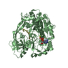
- PDB-7o54: Crystal structure of the carbonic anhydrase-like domain of CcmM i... -
+ Open data

- Basic information
| Entry | Database: PDB / ID: 7o54 | ||||||
|---|---|---|---|---|---|---|---|
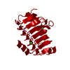
| Title | Crystal structure of the carbonic anhydrase-like domain of CcmM in complex with the C-terminal 17 residues of CcaA from Synechococcus elongatus (strain PCC 7942) | ||||||
 Components Components |
| ||||||
 Keywords Keywords | PHOTOSYNTHESIS / protein binding carboxysome | ||||||
| Function / homology |  Function and homology information Function and homology informationstructural constituent of carboxysome shell / carbon utilization / carboxysome / carbon fixation / photosynthesis / carbonic anhydrase / carbonate dehydratase activity / zinc ion binding Similarity search - Function | ||||||
| Biological species |  Synechococcus elongatus (bacteria) Synechococcus elongatus (bacteria) | ||||||
| Method |  X-RAY DIFFRACTION / SYNCHROTRON / MOLECULAR REPLACEMENT / Resolution: 1.63 Å X-RAY DIFFRACTION / SYNCHROTRON / MOLECULAR REPLACEMENT / Resolution: 1.63 Å | ||||||
 Authors Authors | Zang, K. / Wang, H. / Hartl, F.U. / Hayer-Hartl, M. | ||||||
 Citation Citation | Journal: Nat Struct Mol Biol / Year: 2021 Title: Scaffolding protein CcmM directs multiprotein phase separation in β-carboxysome biogenesis. Authors: Kun Zang / Huping Wang / F Ulrich Hartl / Manajit Hayer-Hartl /  Abstract: Carboxysomes in cyanobacteria enclose the enzymes Rubisco and carbonic anhydrase to optimize photosynthetic carbon fixation. Understanding carboxysome assembly has implications in agricultural ...Carboxysomes in cyanobacteria enclose the enzymes Rubisco and carbonic anhydrase to optimize photosynthetic carbon fixation. Understanding carboxysome assembly has implications in agricultural biotechnology. Here we analyzed the role of the scaffolding protein CcmM of the β-cyanobacterium Synechococcus elongatus PCC 7942 in sequestrating the hexadecameric Rubisco and the tetrameric carbonic anhydrase, CcaA. We find that the trimeric CcmM, consisting of γCAL oligomerization domains and linked small subunit-like (SSUL) modules, plays a central role in mediation of pre-carboxysome condensate formation through multivalent, cooperative interactions. The γCAL domains interact with the C-terminal tails of the CcaA subunits and additionally mediate a head-to-head association of CcmM trimers. Interestingly, SSUL modules, besides their known function in recruiting Rubisco, also participate in intermolecular interactions with the γCAL domains, providing further valency for network formation. Our findings reveal the mechanism by which CcmM functions as a central organizer of the pre-carboxysome multiprotein matrix, concentrating the core components Rubisco and CcaA before β-carboxysome shell formation. | ||||||
| History |
|
- Structure visualization
| Structure viewer | Molecule: MolmilJmol/JSmol |
|---|
- Downloads & links
-Download
| PDBx/mmCIF format | 7o54.cif.gz | 86 KB | Display | PDBx/mmCIF format |
|---|---|---|---|---|
| PDB format | pdb7o54.ent.gz | 63.8 KB | Display | PDB format |
| PDBx/mmJSON format | 7o54.json.gz | Tree view | PDBx/mmJSON format | |
| Others |  Other downloads Other downloads |
-Validation report
| Arichive directory | https://data.pdbj.org/pub/pdb/validation_reports/o5/7o54ftp://data.pdbj.org/pub/pdb/validation_reports/o5/7o54 | HTTPS FTP |
|---|
-Related structure data
| Related structure data |  7o4zSC S: Starting model for refinement C: citing same article ( |
|---|---|
| Similar structure data |


-Links
 PDBj
PDBj


- Assembly
| Deposited unit | 
| |||||||||||||||
|---|---|---|---|---|---|---|---|---|---|---|---|---|---|---|---|---|
| 1 | 
| |||||||||||||||
| Unit cell |
| |||||||||||||||
| Components on special symmetry positions |
|
-Components
| #1: Protein | Mass: 19249.863 Da / Num. of mol.: 1 Source method: isolated from a genetically manipulated source Source: (gene. exp.) Synechococcus elongatus (strain PCC 7942 / FACHB-805) (bacteria)Strain: PCC 7942 / FACHB-805 / Gene: ccmM, Synpcc7942_1423 / Production host: |
|---|---|
| #2: Protein/peptide | Mass: 1976.157 Da / Num. of mol.: 1 Source method: isolated from a genetically manipulated source Source: (gene. exp.) Synechococcus elongatus (strain PCC 7942 / FACHB-805) (bacteria)Strain: PCC 7942 / FACHB-805 / Gene: ccaA, icfA, Synpcc7942_1447 / Production host: |
| #3: Chemical | ChemComp-NI /   Mass: 58.693 Da / Num. of mol.: 1 / Source method: obtained synthetically / Formula: Ni Mass: 58.693 Da / Num. of mol.: 1 / Source method: obtained synthetically / Formula: Ni |
| #4: Chemical | ChemComp-CL /   Mass: 35.453 Da / Num. of mol.: 1 / Source method: obtained synthetically / Formula: Cl Mass: 35.453 Da / Num. of mol.: 1 / Source method: obtained synthetically / Formula: Cl |
| #5: Water | ChemComp-HOH /  Mass: 18.015 Da / Num. of mol.: 103 / Source method: isolated from a natural source / Formula: H2O Mass: 18.015 Da / Num. of mol.: 103 / Source method: isolated from a natural source / Formula: H2O |
| Has ligand of interest | N |
-Experimental details
-Experiment
| Experiment | Method: X-RAY DIFFRACTION / Number of used crystals: 1 |
|---|
- Sample preparation
| Crystal | Density Matthews: 2.32 Å3/Da / Density % sol: 47 % |
|---|---|
| Crystal grow | Temperature: 293 K / Method: vapor diffusion, sitting drop / pH: 7.5 / Details: 25% PEG-3350 and 0.1 M HEPES pH 7.5 |
-Data collection
| Diffraction | Mean temperature: 100 K / Serial crystal experiment: N |
|---|---|
| Diffraction source | Source: SYNCHROTRON / Site: SLS  / Beamline: X06SA / Wavelength: 0.99989 Å / Beamline: X06SA / Wavelength: 0.99989 Å |
| Detector | Type: DECTRIS EIGER X 16M / Detector: PIXEL / Date: Nov 8, 2020 |
| Radiation | Monochromator: M / Protocol: SINGLE WAVELENGTH / Monochromatic (M) / Laue (L): M / Scattering type: x-ray |
| Radiation wavelength | Wavelength: 0.99989 Å / Relative weight: 1 |
| Reflection | Resolution: 1.63→37.08 Å / Num. obs: 24740 / % possible obs: 98.8 % / Redundancy: 18.4 % / CC1/2: 1 / Rmerge(I) obs: 0.03 / Rrim(I) all: 0.031 / Net I/σ(I): 45.4 |
| Reflection shell | Resolution: 1.632→1.661 Å / Rmerge(I) obs: 0.684 / Num. unique obs: 1128 / CC1/2: 0.865 / Rrim(I) all: 0.725 |
- Processing
| Software |
| |||||||||||||||||||||||||||||||||||||||||||||||||||||||||||||||||||||||||||
|---|---|---|---|---|---|---|---|---|---|---|---|---|---|---|---|---|---|---|---|---|---|---|---|---|---|---|---|---|---|---|---|---|---|---|---|---|---|---|---|---|---|---|---|---|---|---|---|---|---|---|---|---|---|---|---|---|---|---|---|---|---|---|---|---|---|---|---|---|---|---|---|---|---|---|---|---|
| Refinement | Method to determine structure: MOLECULAR REPLACEMENT Starting model: 7O4Z Resolution: 1.63→37.08 Å / Cor.coef. Fo:Fc: 0.975 / Cor.coef. Fo:Fc free: 0.972 / SU B: 3.417 / SU ML: 0.057 / Cross valid method: THROUGHOUT / σ(F): 0 / ESU R: 0.086 / ESU R Free: 0.083 / Stereochemistry target values: MAXIMUM LIKELIHOOD Details: HYDROGENS HAVE BEEN ADDED IN THE RIDING POSITIONS U VALUES : WITH TLS ADDED
| |||||||||||||||||||||||||||||||||||||||||||||||||||||||||||||||||||||||||||
| Solvent computation | Ion probe radii: 0.8 Å / Shrinkage radii: 0.8 Å / VDW probe radii: 1.2 Å / Solvent model: MASK | |||||||||||||||||||||||||||||||||||||||||||||||||||||||||||||||||||||||||||
| Displacement parameters | Biso max: 93.71 Å2 / Biso mean: 34.751 Å2 / Biso min: 20.16 Å2
| |||||||||||||||||||||||||||||||||||||||||||||||||||||||||||||||||||||||||||
| Refinement step | Cycle: final / Resolution: 1.63→37.08 Å
| |||||||||||||||||||||||||||||||||||||||||||||||||||||||||||||||||||||||||||
| Refine LS restraints |
| |||||||||||||||||||||||||||||||||||||||||||||||||||||||||||||||||||||||||||
| LS refinement shell | Resolution: 1.632→1.675 Å / Rfactor Rfree error: 0 / Total num. of bins used: 20
| |||||||||||||||||||||||||||||||||||||||||||||||||||||||||||||||||||||||||||
| Refinement TLS params. | Method: refined / Refine-ID: X-RAY DIFFRACTION
| |||||||||||||||||||||||||||||||||||||||||||||||||||||||||||||||||||||||||||
| Refinement TLS group |
|
