 Movie
Movie Controller


+ Open data

- Basic information
| Entry | Database: PDB / ID: 7o4e | ||||||
|---|---|---|---|---|---|---|---|
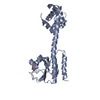
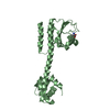
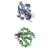
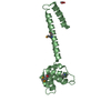
| Title | The DYW domain of A. thaliana OTP86 in its inactive state | ||||||
 Components Components | Pentatricopeptide repeat-containing protein At3g63370, chloroplastic | ||||||
 Keywords Keywords | HYDROLASE / cytidine deaminase | ||||||
| Function / homology |  Function and homology information Function and homology informationchloroplast RNA processing / RNA modification / chloroplast / mRNA processing / mRNA binding / zinc ion binding Similarity search - Function | ||||||
| Biological species |  | ||||||
| Method |  X-RAY DIFFRACTION / SYNCHROTRON / MOLECULAR REPLACEMENT / Resolution: 2.5 Å X-RAY DIFFRACTION / SYNCHROTRON / MOLECULAR REPLACEMENT / Resolution: 2.5 Å | ||||||
 Authors Authors | Weber, G. / Palm, G.J. / Takenaka, M. / Barthel, T. / Feiler, C. / Weiss, M.S. | ||||||
 Citation Citation | Journal: Nat Catal / Year: 2021 Title: DYW domain structures imply an unusual regulation principle in plant organellar RNA editing catalysis. Authors: Takenaka, M. / Takenaka, S. / Barthel, T. / Frink, B. / Haag, S. / Verbitskiy, D. / Oldenkott, B. / Schallenberg-Rudinger, M. / Feiler, C.G. / Weiss, M.S. / Palm, G.J. / Weber, G. | ||||||
| History |
|
- Structure visualization
| Structure viewer | Molecule: MolmilJmol/JSmol |
|---|
- Downloads & links
-Download
| PDBx/mmCIF format | 7o4e.cif.gz | 67.2 KB | Display | PDBx/mmCIF format |
|---|---|---|---|---|
| PDB format | pdb7o4e.ent.gz | 48.5 KB | Display | PDB format |
| PDBx/mmJSON format | 7o4e.json.gz | Tree view | PDBx/mmJSON format | |
| Others |  Other downloads Other downloads |
-Validation report
| Summary document | 7o4e_validation.pdf.gz | 439.9 KB | Display | wwPDB validaton report |
|---|---|---|---|---|
| Full document | 7o4e_full_validation.pdf.gz | 441.9 KB | Display | |
| Data in XML | 7o4e_validation.xml.gz | 11.5 KB | Display | |
| Data in CIF | 7o4e_validation.cif.gz | 14.7 KB | Display | |
| Arichive directory | https://data.pdbj.org/pub/pdb/validation_reports/o4/7o4eftp://data.pdbj.org/pub/pdb/validation_reports/o4/7o4e | HTTPS FTP |
-Related structure data


-Links
 PDBj
PDBj
- Assembly
| Deposited unit | 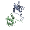
| |||||||||
|---|---|---|---|---|---|---|---|---|---|---|
| 1 | 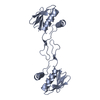
| |||||||||
| 2 | 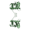
| |||||||||
| Unit cell |
| |||||||||
| Components on special symmetry positions |
|

-Components
| #1: Protein | Mass: 16016.490 Da / Num. of mol.: 2 Source method: isolated from a genetically manipulated source Source: (gene. exp.)  #2: Chemical | ChemComp-ZN /   Mass: 65.409 Da / Num. of mol.: 4 / Source method: obtained synthetically / Formula: Zn Mass: 65.409 Da / Num. of mol.: 4 / Source method: obtained synthetically / Formula: Zn#3: Water | ChemComp-HOH / |  Mass: 18.015 Da / Num. of mol.: 33 / Source method: isolated from a natural source / Formula: H2O Mass: 18.015 Da / Num. of mol.: 33 / Source method: isolated from a natural source / Formula: H2OHas ligand of interest | N | |
|---|
-Experimental details
-Experiment
| Experiment | Method: X-RAY DIFFRACTION / Number of used crystals: 1 |
|---|
- Sample preparation
| Crystal | Density Matthews: 1.92 Å3/Da / Density % sol: 36 % |
|---|---|
| Crystal grow | Temperature: 277 K / Method: vapor diffusion, sitting drop Details: 100 nl protein plus 100 nl reservoir (0.1 M glycine, pH 10.5, 1.2 M NaH2PO4, 0.8 M K2HPO4, 0.2 M Li2SO4) plus 30 nl additive (0.1 M 50% v/v Jeffamine M-600 pH 7.0). Cryo: reservoir solution ...Details: 100 nl protein plus 100 nl reservoir (0.1 M glycine, pH 10.5, 1.2 M NaH2PO4, 0.8 M K2HPO4, 0.2 M Li2SO4) plus 30 nl additive (0.1 M 50% v/v Jeffamine M-600 pH 7.0). Cryo: reservoir solution supplemented with 15% (v/v) ethylene glycol. PH range: 7.9 - 10.5 |
-Data collection
| Diffraction | Mean temperature: 100 K / Serial crystal experiment: N |
|---|---|
| Diffraction source | Source: SYNCHROTRON / Site: BESSY  / Beamline: 14.1 / Wavelength: 1.282 Å / Beamline: 14.1 / Wavelength: 1.282 Å |
| Detector | Type: DECTRIS PILATUS 6M / Detector: PIXEL / Date: May 15, 2018 |
| Radiation | Protocol: SINGLE WAVELENGTH / Monochromatic (M) / Laue (L): M / Scattering type: x-ray |
| Radiation wavelength | Wavelength: 1.282 Å / Relative weight: 1 |
| Reflection | Resolution: 2.5→80 Å / Num. obs: 16019 / % possible obs: 99.2 % / Redundancy: 6.5 % / Biso Wilson estimate: 48.3 Å2 / CC1/2: 0.994 / Rrim(I) all: 0.235 / Rsym value: 0.216 / Net I/σ(I): 7.46 |
| Reflection shell | Resolution: 2.5→2.6 Å / Redundancy: 6 % / Mean I/σ(I) obs: 1.06 / Num. unique obs: 1761 / CC1/2: 0.428 / Rrim(I) all: 1.899 / Rsym value: 1.734 / % possible all: 99 |
- Processing
| Software |
| ||||||||||||||||||||||||
|---|---|---|---|---|---|---|---|---|---|---|---|---|---|---|---|---|---|---|---|---|---|---|---|---|---|
| Refinement | Method to determine structure: MOLECULAR REPLACEMENT Starting model: D_129211461 Resolution: 2.5→38.722 Å / SU ML: 0.41 / Cross valid method: THROUGHOUT / σ(F): 1.36 / Phase error: 32.54 / Stereochemistry target values: ML
| ||||||||||||||||||||||||
| Solvent computation | Shrinkage radii: 0.9 Å / VDW probe radii: 1.11 Å / Solvent model: FLAT BULK SOLVENT MODEL | ||||||||||||||||||||||||
| Displacement parameters | Biso max: 96.8 Å2 / Biso mean: 48.7795 Å2 / Biso min: 26.76 Å2 | ||||||||||||||||||||||||
| Refinement step | Cycle: final / Resolution: 2.5→38.722 Å
| ||||||||||||||||||||||||
| LS refinement shell | Refine-ID: X-RAY DIFFRACTION / Rfactor Rfree error: 0
|
