 Movie
Movie Controller


+ Open data

- Basic information
| Entry | Database: PDB / ID: 7nyo | |||||||||
|---|---|---|---|---|---|---|---|---|---|---|
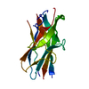
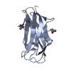
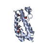
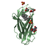
| Title | Mutant A541L of SH3 domain of JNK-interacting Protein 1 (JIP1) | |||||||||
 Components Components | SH3 domain of JNK-interacting Protein 1 (JIP1) | |||||||||
 Keywords Keywords | SIGNALING PROTEIN / SH3 domain of JNK-interacting protein 1 (JIP1) | |||||||||
| Function / homology |  Function and homology information Function and homology informationdentate gyrus mossy fiber / regulation of CD8-positive, alpha-beta T cell proliferation / negative regulation of JUN kinase activity / MAP kinase scaffold activity / JUN kinase binding / mitogen-activated protein kinase kinase binding / mitogen-activated protein kinase kinase kinase binding / regulation of JNK cascade / dendritic growth cone / protein kinase inhibitor activity ...dentate gyrus mossy fiber / regulation of CD8-positive, alpha-beta T cell proliferation / negative regulation of JUN kinase activity / MAP kinase scaffold activity / JUN kinase binding / mitogen-activated protein kinase kinase binding / mitogen-activated protein kinase kinase kinase binding / regulation of JNK cascade / dendritic growth cone / protein kinase inhibitor activity / JNK cascade / kinesin binding / axonal growth cone / vesicle-mediated transport / positive regulation of JNK cascade / mitochondrial membrane / cell body / synapse / negative regulation of apoptotic process / endoplasmic reticulum membrane / perinuclear region of cytoplasm / nucleus / plasma membrane / cytosol / cytoplasm Similarity search - Function | |||||||||
| Biological species |  Homo sapiens (human) Homo sapiens (human) | |||||||||
| Method |  X-RAY DIFFRACTION / SYNCHROTRON / MOLECULAR REPLACEMENT / Resolution: 1.4 Å X-RAY DIFFRACTION / SYNCHROTRON / MOLECULAR REPLACEMENT / Resolution: 1.4 Å | |||||||||
 Authors Authors | Perez, L.M. / Ielasi, F.S. / Palencia, A. / Jensen, M.R. | |||||||||
| Funding support |  France, 2items France, 2items
| |||||||||
 Citation Citation | Journal: Nature / Year: 2022 Title: Visualizing protein breathing motions associated with aromatic ring flipping. Authors: Marino Perez, L. / Ielasi, F.S. / Bessa, L.M. / Maurin, D. / Kragelj, J. / Blackledge, M. / Salvi, N. / Bouvignies, G. / Palencia, A. / Jensen, M.R. | |||||||||
| History |
|
- Structure visualization
| Structure viewer | Molecule: MolmilJmol/JSmol |
|---|
- Downloads & links
-Download
| PDBx/mmCIF format | 7nyo.cif.gz | 135.3 KB | Display | PDBx/mmCIF format |
|---|---|---|---|---|
| PDB format | pdb7nyo.ent.gz | Display | PDB format | |
| PDBx/mmJSON format | 7nyo.json.gz | Tree view | PDBx/mmJSON format | |
| Others |  Other downloads Other downloads |
-Validation report
| Arichive directory | https://data.pdbj.org/pub/pdb/validation_reports/ny/7nyoftp://data.pdbj.org/pub/pdb/validation_reports/ny/7nyo | HTTPS FTP |
|---|
-Related structure data
| Related structure data |  7nykSC  7nylC  7nymC  7nynC  7nzbC  7nzcC  7nzdC S: Starting model for refinement C: citing same article ( |
|---|---|
| Similar structure data |


-Links
 PDBj
PDBj
- Assembly
| Deposited unit | 
| |||||||||||||||
|---|---|---|---|---|---|---|---|---|---|---|---|---|---|---|---|---|
| 1 | 
| |||||||||||||||
| 2 | 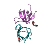
| |||||||||||||||
| Unit cell |
| |||||||||||||||
| Components on special symmetry positions |
|
-Components
| #1: Protein | Mass: 7568.447 Da / Num. of mol.: 4 / Mutation: A541L Source method: isolated from a genetically manipulated source Details: GHM belongs to expression vector pET28a / Source: (gene. exp.) Homo sapiens (human) / Gene: MAPK8IP1, IB1, JIP1, PRKM8IP / Production host:  #2: Chemical |   Mass: 194.226 Da / Num. of mol.: 3 / Source method: obtained synthetically / Formula: C8H18O5 / Comment: precipitant*YM Mass: 194.226 Da / Num. of mol.: 3 / Source method: obtained synthetically / Formula: C8H18O5 / Comment: precipitant*YM#3: Chemical | ChemComp-SO4 / |   Mass: 96.063 Da / Num. of mol.: 1 / Source method: obtained synthetically / Formula: SO4 Mass: 96.063 Da / Num. of mol.: 1 / Source method: obtained synthetically / Formula: SO4#4: Chemical | ChemComp-EDO / |   Mass: 62.068 Da / Num. of mol.: 1 / Source method: obtained synthetically / Formula: C2H6O2 Mass: 62.068 Da / Num. of mol.: 1 / Source method: obtained synthetically / Formula: C2H6O2#5: Water | ChemComp-HOH / |  Mass: 18.015 Da / Num. of mol.: 525 / Source method: isolated from a natural source / Formula: H2O Mass: 18.015 Da / Num. of mol.: 525 / Source method: isolated from a natural source / Formula: H2OHas ligand of interest | N | |
|---|
-Experimental details
-Experiment
| Experiment | Method: X-RAY DIFFRACTION / Number of used crystals: 1 |
|---|
- Sample preparation
| Crystal | Density Matthews: 2 Å3/Da / Density % sol: 40.5 % / Description: 3D needles |
|---|---|
| Crystal grow | Temperature: 293 K / Method: vapor diffusion / pH: 7.5 Details: 0.1 M HEPES pH 7.5, 1-5 % PEG 400 and 2-2.5 M ammonium sulfate |
-Data collection
| Diffraction | Mean temperature: 100 K / Serial crystal experiment: N |
|---|---|
| Diffraction source | Source: SYNCHROTRON / Site: ESRF / Beamline: ID23-2 / Wavelength: 0.873128 Å |
| Detector | Type: DECTRIS PILATUS 6M / Detector: PIXEL / Date: Oct 30, 2020 |
| Radiation | Monochromator: SILICON CRYSTAL / Protocol: SINGLE WAVELENGTH / Monochromatic (M) / Laue (L): M / Scattering type: x-ray |
| Radiation wavelength | Wavelength: 0.873128 Å / Relative weight: 1 |
| Reflection | Resolution: 1.4→47.418 Å / Num. obs: 41884 / % possible obs: 91.9 % / Redundancy: 5.3 % / CC1/2: 0.982 / Net I/σ(I): 5.5 |
| Reflection shell | Resolution: 1.4→1.51 Å / Mean I/σ(I) obs: 1.5 / Num. unique obs: 2095 / CC1/2: 0.74 |
- Processing
| Software |
| ||||||||||||||||||||||||||||||||||||||||||||||||||||||||||||||||||||||||||||||||||||||||||||||||||||||||||||||||||||||||||||||||||||||||||||||||||||||||||||||||
|---|---|---|---|---|---|---|---|---|---|---|---|---|---|---|---|---|---|---|---|---|---|---|---|---|---|---|---|---|---|---|---|---|---|---|---|---|---|---|---|---|---|---|---|---|---|---|---|---|---|---|---|---|---|---|---|---|---|---|---|---|---|---|---|---|---|---|---|---|---|---|---|---|---|---|---|---|---|---|---|---|---|---|---|---|---|---|---|---|---|---|---|---|---|---|---|---|---|---|---|---|---|---|---|---|---|---|---|---|---|---|---|---|---|---|---|---|---|---|---|---|---|---|---|---|---|---|---|---|---|---|---|---|---|---|---|---|---|---|---|---|---|---|---|---|---|---|---|---|---|---|---|---|---|---|---|---|---|---|---|---|---|
| Refinement | Method to determine structure: MOLECULAR REPLACEMENT Starting model: 7NYK Resolution: 1.4→47.418 Å / Cor.coef. Fo:Fc: 0.957 / Cor.coef. Fo:Fc free: 0.908 / SU B: 5.389 / SU ML: 0.093 / Cross valid method: FREE R-VALUE / ESU R: 0.126 / ESU R Free: 0.114 Details: Hydrogens have been added in their riding positions
| ||||||||||||||||||||||||||||||||||||||||||||||||||||||||||||||||||||||||||||||||||||||||||||||||||||||||||||||||||||||||||||||||||||||||||||||||||||||||||||||||
| Solvent computation | Ion probe radii: 0.8 Å / Shrinkage radii: 0.8 Å / VDW probe radii: 1.2 Å / Solvent model: MASK BULK SOLVENT | ||||||||||||||||||||||||||||||||||||||||||||||||||||||||||||||||||||||||||||||||||||||||||||||||||||||||||||||||||||||||||||||||||||||||||||||||||||||||||||||||
| Displacement parameters | Biso mean: 23.522 Å2
| ||||||||||||||||||||||||||||||||||||||||||||||||||||||||||||||||||||||||||||||||||||||||||||||||||||||||||||||||||||||||||||||||||||||||||||||||||||||||||||||||
| Refinement step | Cycle: LAST / Resolution: 1.4→47.418 Å
| ||||||||||||||||||||||||||||||||||||||||||||||||||||||||||||||||||||||||||||||||||||||||||||||||||||||||||||||||||||||||||||||||||||||||||||||||||||||||||||||||
| Refine LS restraints |
| ||||||||||||||||||||||||||||||||||||||||||||||||||||||||||||||||||||||||||||||||||||||||||||||||||||||||||||||||||||||||||||||||||||||||||||||||||||||||||||||||
| LS refinement shell |
|
