[English] 日本語
Yorodumi
- PDB-7nxm: Structure of human cathepsin K in complex with the selective acti... -

+
Open data


ID or keywords:

Loading...

-
Basic information

Entry
Database: PDB / ID: 7nxm
TitleStructure of human cathepsin K in complex with the selective activity-based probe Gu3416
ComponentsCathepsin K
KeywordsHYDROLASE / inhibitor / complex / activity-based probe / acrylamide inhibitor
Function / homology
Function and homology information


cathepsin K / mononuclear cell differentiation / intramembranous ossification / negative regulation of cartilage development / cellular response to zinc ion starvation / RUNX1 regulates transcription of genes involved in differentiation of keratinocytes / thyroid hormone generation / endolysosome lumen / Trafficking and processing of endosomal TLR / proteoglycan binding ...cathepsin K / mononuclear cell differentiation / intramembranous ossification / negative regulation of cartilage development / cellular response to zinc ion starvation / RUNX1 regulates transcription of genes involved in differentiation of keratinocytes / thyroid hormone generation / endolysosome lumen / Trafficking and processing of endosomal TLR / proteoglycan binding / Activation of Matrix Metalloproteinases / cysteine-type endopeptidase activator activity involved in apoptotic process / mitophagy / fibronectin binding / Collagen degradation / collagen catabolic process / extracellular matrix disassembly / cysteine-type peptidase activity / bone resorption / cellular response to transforming growth factor beta stimulus / collagen binding / MHC class II antigen presentation / Degradation of the extracellular matrix / lysosomal lumen / proteolysis involved in protein catabolic process / positive regulation of apoptotic signaling pathway / response to insulin / response to organic cyclic compound / cellular response to tumor necrosis factor / response to ethanol / lysosome / immune response / apical plasma membrane / external side of plasma membrane / cysteine-type endopeptidase activity / intracellular membrane-bounded organelle / serine-type endopeptidase activity / proteolysis / extracellular space / extracellular region / nucleoplasm
Similarity search - Function
Cathepsin propeptide inhibitor domain (I29) / Cathepsin propeptide inhibitor domain (I29) / Cathepsin propeptide inhibitor domain (I29) / Papain-like cysteine endopeptidase / : / Cysteine peptidase, asparagine active site / Eukaryotic thiol (cysteine) proteases asparagine active site. / Cysteine peptidase, histidine active site / Eukaryotic thiol (cysteine) proteases histidine active site. / Peptidase C1A, papain C-terminal ...Cathepsin propeptide inhibitor domain (I29) / Cathepsin propeptide inhibitor domain (I29) / Cathepsin propeptide inhibitor domain (I29) / Papain-like cysteine endopeptidase / : / Cysteine peptidase, asparagine active site / Eukaryotic thiol (cysteine) proteases asparagine active site. / Cysteine peptidase, histidine active site / Eukaryotic thiol (cysteine) proteases histidine active site. / Peptidase C1A, papain C-terminal / Papain family cysteine protease / Papain family cysteine protease / Cysteine peptidase, cysteine active site / Eukaryotic thiol (cysteine) proteases cysteine active site. / Papain-like cysteine peptidase superfamily
Similarity search - Domain/homology
Chem-UUW / Cathepsin K
Similarity search - Component
Biological speciesHomo sapiens (human)
MethodX-RAY DIFFRACTION / MOLECULAR REPLACEMENT / Resolution: 1.72 Å
AuthorsBusa, M. / Benysek, J. / Lemke, C. / Gutschow, M. / Mares, M.
CitationJournal: J.Med.Chem. / Year: 2021
Title: An Activity-Based Probe for Cathepsin K Imaging with Excellent Potency and Selectivity.
Authors: Lemke, C. / Benysek, J. / Brajtenbach, D. / Breuer, C. / Jilkova, A. / Horn, M. / Busa, M. / Ulrychova, L. / Illies, A. / Kubatzky, K.F. / Bartz, U. / Mares, M. / Gutschow, M.
History
DepositionMar 18, 2021Deposition site: PDBE / Processing site: PDBE
Revision 1.0Sep 8, 2021Provider: repository / Type: Initial release
Revision 1.1Oct 13, 2021Group: Data collection / Database references / Category: citation / citation_author / pdbx_database_proc
Item: _citation.journal_volume / _citation.page_first ..._citation.journal_volume / _citation.page_first / _citation.page_last / _citation.pdbx_database_id_DOI / _citation.pdbx_database_id_PubMed / _citation.title
Revision 1.2Jan 31, 2024Group: Data collection / Refinement description
Category: chem_comp_atom / chem_comp_bond / pdbx_initial_refinement_model

-
Structure visualization

Structure viewerMolecule:
MolmilJmol/JSmol

Downloads & links

-
Assembly

Deposited unit
A: Cathepsin K
hetero molecules


Theoretical massNumber of molelcules
Total (without water)24,2993
Polymers23,6811
Non-polymers6192
Water3,315184
1


  • Idetical with deposited unit
  • defined by author&software
  • Evidence: gel filtration
TypeNameSymmetry operationNumber
identity operation1_555x,y,z1
Buried area150 Å2
ΔGint-8 kcal/mol
Surface area9850 Å2
MethodPISA
Unit cell
Length a, b, c (Å)32.045, 71.677, 80.856
Angle α, β, γ (deg.)90.000, 90.000, 90.000
Int Tables number18
Space group name H-MP22121

-
Components

#1: Protein Cathepsin K / Cathepsin O / Cathepsin O2 / Cathepsin X


Mass: 23680.674 Da / Num. of mol.: 1
Source method: isolated from a genetically manipulated source
Source: (gene. exp.) Homo sapiens (human) / Gene: CTSK, CTSO, CTSO2 / Production host: 
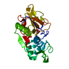
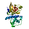
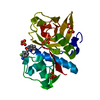
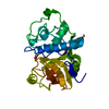
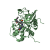
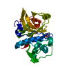
Komagataella phaffii GS115 (fungus) / References: UniProt: P43235, cathepsin K
#2: Chemical ChemComp-SO4 / SULFATE ION


Mass: 96.063 Da / Num. of mol.: 1 / Source method: obtained synthetically / Formula: SO4
#3: Chemical ChemComp-UUW / N-(4-(dibenzylamino)-4-oxobutyl)-2-(5-(dimethylamino)pentanamido)-4-methylpentanamide / (2~{S})-~{N}-[4-[bis(phenylmethyl)amino]-4-oxidanylidene-butyl]-2-[5-(dimethylamino)pentanoylamino]-4-methyl-pentanamide / activity-based probe Gu3416 / (2S)-N-[4-[bis(phenylmethyl)amino]-4-oxidanylidene-butyl]-2-[5-(dimethylamino)pentanoylamino]-4-methyl-pentanamide


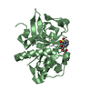
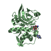
Mass: 522.722 Da / Num. of mol.: 1 / Source method: obtained synthetically / Formula: C31H46N4O3 / Feature type: SUBJECT OF INVESTIGATION
#4: Water ChemComp-HOH / water


Mass: 18.015 Da / Num. of mol.: 184 / Source method: isolated from a natural source / Formula: H2O
Has ligand of interestY

-
Experimental details

-
Experiment

ExperimentMethod: X-RAY DIFFRACTION / Number of used crystals: 1

-
Sample preparation

CrystalDensity Matthews: 1.963 Å3/Da / Density % sol: 37.38 % / Description: Orthorhombic crystals
Crystal growTemperature: 291 K / Method: vapor diffusion, hanging drop / pH: 6.5
Details: Composition of reservoir solution was 0.1 M MES/imidazole buffer, containing: 12.5% w/v PEG 1000, 12.5% w/v PEG 3350, 12.5% v/v MPD 0.03 M of each NPS (a mix containing sodium nitrate, ...Details: Composition of reservoir solution was 0.1 M MES/imidazole buffer, containing: 12.5% w/v PEG 1000, 12.5% w/v PEG 3350, 12.5% v/v MPD 0.03 M of each NPS (a mix containing sodium nitrate, disodium hydrogen phosphate and ammonium sulfate) additives, pH 6.5. Morpheus C4 condition

-
Data collection

DiffractionMean temperature: 100 K / Serial crystal experiment: N
Diffraction sourceSource: ROTATING ANODE / Type: RIGAKU MICROMAX-007 / Wavelength: 1.54187 Å
DetectorType: DECTRIS PILATUS 300K / Detector: PIXEL / Date: Mar 5, 2020
RadiationMonochromator: M / Protocol: SINGLE WAVELENGTH / Monochromatic (M) / Laue (L): M / Scattering type: x-ray
Radiation wavelengthWavelength: 1.54187 Å / Relative weight: 1
ReflectionResolution: 1.72→50 Å / Num. obs: 17548 / % possible obs: 85.7 % / Redundancy: 3.8 % / CC1/2: 0.996 / Rmerge(I) obs: 0.061 / Rpim(I) all: 0.05 / Rrim(I) all: 0.079 / Χ2: 0.89 / Net I/av σ(I): 15.81 / Net I/σ(I): 27.18
Reflection shell

Diffraction-ID: 1

Resolution (Å)Redundancy (%)Rmerge(I) obsMean I/σ(I) obsNum. unique obsCC1/2Rpim(I) allRrim(I) allΧ2% possible all
1.72-1.751.30.4231.41530.6780.4230.5980.5814.8
1.8-1.842.80.2863.17720.8810.2740.3970.7674.9

-
Processing

Software
NameVersionClassification
REFMAC5.8.0267refinement
HKL-3000data reduction
HKL-3000data scaling
PDB_EXTRACT3.27data extraction
MOLREPphasing
RefinementMethod to determine structure: MOLECULAR REPLACEMENT
Starting model: 5TDI
Resolution: 1.72→35.86 Å / Cor.coef. Fo:Fc: 0.961 / Cor.coef. Fo:Fc free: 0.951 / SU B: 2.167 / SU ML: 0.069 / Cross valid method: THROUGHOUT / ESU R: 0.137 / ESU R Free: 0.112
Details: Hydrogens have been added in their riding positions
RfactorNum. reflection% reflectionSelection details
Rfree0.1784 895 5.111 %RANDOM
Rwork0.1602 16615 --
all0.161 ---
obs-17510 85.34 %-
Solvent computationIon probe radii: 0.8 Å / Shrinkage radii: 0.8 Å / VDW probe radii: 1.2 Å / Solvent model: MASK BULK SOLVENT
Displacement parametersBiso mean: 17.819 Å2
Baniso -1Baniso -2Baniso -3
1-0.356 Å2-0 Å2-0 Å2
2---1.202 Å20 Å2
3---0.846 Å2
Refinement stepCycle: LAST / Resolution: 1.72→35.86 Å
ProteinNucleic acidLigandSolventTotal
Num. atoms1652 0 37 184 1873
Refine LS restraints
Refine-IDTypeDev idealDev ideal targetNumber
X-RAY DIFFRACTIONr_bond_refined_d0.0120.0131776
X-RAY DIFFRACTIONr_bond_other_d0.0020.0181640
X-RAY DIFFRACTIONr_angle_refined_deg1.6331.642408
X-RAY DIFFRACTIONr_angle_other_deg1.4651.5893778
X-RAY DIFFRACTIONr_dihedral_angle_1_deg5.8965231
X-RAY DIFFRACTIONr_dihedral_angle_2_deg35.74622.63295
X-RAY DIFFRACTIONr_dihedral_angle_3_deg12.42315293
X-RAY DIFFRACTIONr_dihedral_angle_other_3_deg13.112151
X-RAY DIFFRACTIONr_dihedral_angle_4_deg17.2251510
X-RAY DIFFRACTIONr_chiral_restr0.0850.2215
X-RAY DIFFRACTIONr_gen_planes_refined0.0090.022104
X-RAY DIFFRACTIONr_gen_planes_other0.0010.02440
X-RAY DIFFRACTIONr_nbd_refined0.2090.2349
X-RAY DIFFRACTIONr_symmetry_nbd_other0.1890.21535
X-RAY DIFFRACTIONr_nbtor_refined0.1750.2871
X-RAY DIFFRACTIONr_symmetry_nbtor_other0.0840.2775
X-RAY DIFFRACTIONr_xyhbond_nbd_refined0.1530.2130
X-RAY DIFFRACTIONr_symmetry_nbd_refined0.1410.27
X-RAY DIFFRACTIONr_nbd_other0.1670.237
X-RAY DIFFRACTIONr_symmetry_xyhbond_nbd_refined0.2230.27
X-RAY DIFFRACTIONr_mcbond_it1.6751.598879
X-RAY DIFFRACTIONr_mcbond_other1.6611.596878
X-RAY DIFFRACTIONr_mcangle_it2.3772.3871101
X-RAY DIFFRACTIONr_mcangle_other2.382.3891102
X-RAY DIFFRACTIONr_scbond_it2.5792.014897
X-RAY DIFFRACTIONr_scbond_other2.5782.016898
X-RAY DIFFRACTIONr_scangle_it3.852.881299
X-RAY DIFFRACTIONr_scangle_other3.8492.8821300
X-RAY DIFFRACTIONr_lrange_it5.2819.9762065
X-RAY DIFFRACTIONr_lrange_other5.1419.7452043
LS refinement shell
Resolution (Å)Rfactor RfreeNum. reflection RfreeRfactor RworkNum. reflection RworkRefine-ID% reflection obs (%)
1.72-1.7650.409200.256272X-RAY DIFFRACTION19.6369
1.765-1.8130.241460.247771X-RAY DIFFRACTION56.9338
1.813-1.8660.267660.2031050X-RAY DIFFRACTION78.4259
1.866-1.9230.174680.1751078X-RAY DIFFRACTION84.0176
1.923-1.9860.143470.1511107X-RAY DIFFRACTION86.5716
1.986-2.0560.214670.1611113X-RAY DIFFRACTION91.2606
2.056-2.1330.137610.1451118X-RAY DIFFRACTION93.2016
2.133-2.220.16540.1361104X-RAY DIFFRACTION98.2188
2.22-2.3190.124650.1381065X-RAY DIFFRACTION96.7466
2.319-2.4320.204650.151023X-RAY DIFFRACTION99.2701
2.432-2.5630.155480.154991X-RAY DIFFRACTION97.2846
2.563-2.7190.165420.167943X-RAY DIFFRACTION97.0443
2.719-2.9060.234440.165889X-RAY DIFFRACTION98.5217
2.906-3.1390.175430.152817X-RAY DIFFRACTION98.1735
3.139-3.4380.255350.164763X-RAY DIFFRACTION98.2759
3.438-3.8430.145420.148678X-RAY DIFFRACTION94.9868
3.843-4.4350.144310.14563X-RAY DIFFRACTION89.1892
4.435-5.4280.2220.164564X-RAY DIFFRACTION100
5.428-7.6590.186180.203435X-RAY DIFFRACTION100
7.659-35.860.145110.18271X-RAY DIFFRACTION98.9474

+
About Yorodumi

-
News

-
Feb 9, 2022. New format data for meta-information of EMDB entries

New format data for meta-information of EMDB entries

  • Version 3 of the EMDB header file is now the official format.
  • The previous official version 1.9 will be removed from the archive.

Related info.:EMDB header

External links:wwPDB to switch to version 3 of the EMDB data model

-
Aug 12, 2020. Covid-19 info

Covid-19 info

URL: https://pdbjlvh1.pdbj.org/emnavi/covid19.php

New page: Covid-19 featured information page in EM Navigator.

Related info.:Covid-19 info / Mar 5, 2020. Novel coronavirus structure data

+
Mar 5, 2020. Novel coronavirus structure data

Novel coronavirus structure data

Related info.:Yorodumi Speices / Aug 12, 2020. Covid-19 info

External links:COVID-19 featured content - PDBj / Molecule of the Month (242):Coronavirus Proteases

+
Jan 31, 2019. EMDB accession codes are about to change! (news from PDBe EMDB page)

EMDB accession codes are about to change! (news from PDBe EMDB page)

  • The allocation of 4 digits for EMDB accession codes will soon come to an end. Whilst these codes will remain in use, new EMDB accession codes will include an additional digit and will expand incrementally as the available range of codes is exhausted. The current 4-digit format prefixed with “EMD-” (i.e. EMD-XXXX) will advance to a 5-digit format (i.e. EMD-XXXXX), and so on. It is currently estimated that the 4-digit codes will be depleted around Spring 2019, at which point the 5-digit format will come into force.
  • The EM Navigator/Yorodumi systems omit the EMD- prefix.

Related info.:Q: What is EMD? / ID/Accession-code notation in Yorodumi/EM Navigator

External links:EMDB Accession Codes are Changing Soon! / Contact to PDBj

+
Jul 12, 2017. Major update of PDB

Major update of PDB

  • wwPDB released updated PDB data conforming to the new PDBx/mmCIF dictionary.
  • This is a major update changing the version number from 4 to 5, and with Remediation, in which all the entries are updated.
  • In this update, many items about electron microscopy experimental information are reorganized (e.g. em_software).
  • Now, EM Navigator and Yorodumi are based on the updated data.

External links:wwPDB Remediation / Enriched Model Files Conforming to OneDep Data Standards Now Available in the PDB FTP Archive

-
Yorodumi

Thousand views of thousand structures

  • Yorodumi is a browser for structure data from EMDB, PDB, SASBDB, etc.
  • This page is also the successor to EM Navigator detail page, and also detail information page/front-end page for Omokage search.
  • The word "yorodu" (or yorozu) is an old Japanese word meaning "ten thousand". "mi" (miru) is to see.

Related info.:EMDB / PDB / SASBDB / Comparison of 3 databanks / Yorodumi Search / Aug 31, 2016. New EM Navigator & Yorodumi / Yorodumi Papers / Jmol/JSmol / Function and homology information / Changes in new EM Navigator and Yorodumi

Read more