 Movie
Movie Controller

[English] 日本語
 Yorodumi
- PDB-7nud: Crystal structure of mouse PRMT6 in complex with inhibitor EML734 -
+ Open data

- Basic information
| Entry | Database: PDB / ID: 7nud | |||||||||||||||
|---|---|---|---|---|---|---|---|---|---|---|---|---|---|---|---|---|
| Title | Crystal structure of mouse PRMT6 in complex with inhibitor EML734 | |||||||||||||||
 Components Components | Protein arginine N-methyltransferase 6 | |||||||||||||||
 Keywords Keywords | TRANSFERASE / SAM binding domain / arginine methylation | |||||||||||||||
| Function / homology |  Function and homology information Function and homology informationhistone H2AR3 methyltransferase activity / protein-arginine omega-N monomethyltransferase activity / RUNX1 regulates genes involved in megakaryocyte differentiation and platelet function / histone H3R2 methyltransferase activity / RMTs methylate histone arginines / protein-arginine omega-N asymmetric methyltransferase activity / type I protein arginine methyltransferase / histone H4R3 methyltransferase activity / : / protein-arginine N-methyltransferase activity ...histone H2AR3 methyltransferase activity / protein-arginine omega-N monomethyltransferase activity / RUNX1 regulates genes involved in megakaryocyte differentiation and platelet function / histone H3R2 methyltransferase activity / RMTs methylate histone arginines / protein-arginine omega-N asymmetric methyltransferase activity / type I protein arginine methyltransferase / histone H4R3 methyltransferase activity / : / protein-arginine N-methyltransferase activity / regulation of mitochondrion organization / histone methyltransferase activity / negative regulation of ubiquitin-dependent protein catabolic process / regulation of signal transduction by p53 class mediator / protein modification process / cellular senescence / histone binding / methylation / DNA repair / negative regulation of DNA-templated transcription / chromatin binding / nucleolus / negative regulation of transcription by RNA polymerase II / nucleoplasm / nucleus Similarity search - Function | |||||||||||||||
| Biological species |  | |||||||||||||||
| Method |  X-RAY DIFFRACTION / MOLECULAR REPLACEMENT / molecular replacement / Resolution: 1.65 Å X-RAY DIFFRACTION / MOLECULAR REPLACEMENT / molecular replacement / Resolution: 1.65 Å | |||||||||||||||
 Authors Authors | Bonnefond, L. / Cavarelli, J. | |||||||||||||||
| Funding support |  France, 4items France, 4items
| |||||||||||||||
 Citation Citation | Journal: J.Med.Chem. / Year: 2022 Title: Turning Nonselective Inhibitors of Type I Protein Arginine Methyltransferases into Potent and Selective Inhibitors of Protein Arginine Methyltransferase 4 through a Deconstruction- ...Title: Turning Nonselective Inhibitors of Type I Protein Arginine Methyltransferases into Potent and Selective Inhibitors of Protein Arginine Methyltransferase 4 through a Deconstruction-Reconstruction and Fragment-Growing Approach. Authors: Iannelli, G. / Milite, C. / Marechal, N. / Cura, V. / Bonnefond, L. / Troffer-Charlier, N. / Feoli, A. / Rescigno, D. / Wang, Y. / Cipriano, A. / Viviano, M. / Bedford, M.T. / Cavarelli, J. ...Authors: Iannelli, G. / Milite, C. / Marechal, N. / Cura, V. / Bonnefond, L. / Troffer-Charlier, N. / Feoli, A. / Rescigno, D. / Wang, Y. / Cipriano, A. / Viviano, M. / Bedford, M.T. / Cavarelli, J. / Castellano, S. / Sbardella, G. | |||||||||||||||
| History |
|
- Structure visualization
| Structure viewer | Molecule: MolmilJmol/JSmol |
|---|
- Downloads & links
-Download
| PDBx/mmCIF format | 7nud.cif.gz | 377.1 KB | Display | PDBx/mmCIF format |
|---|---|---|---|---|
| PDB format | pdb7nud.ent.gz | 313.7 KB | Display | PDB format |
| PDBx/mmJSON format | 7nud.json.gz | Tree view | PDBx/mmJSON format | |
| Others |  Other downloads Other downloads |
-Validation report
| Summary document | 7nud_validation.pdf.gz | 1.1 MB | Display | wwPDB validaton report |
|---|---|---|---|---|
| Full document | 7nud_full_validation.pdf.gz | 1.1 MB | Display | |
| Data in XML | 7nud_validation.xml.gz | 25.2 KB | Display | |
| Data in CIF | 7nud_validation.cif.gz | 35.5 KB | Display | |
| Arichive directory | https://data.pdbj.org/pub/pdb/validation_reports/nu/7nudftp://data.pdbj.org/pub/pdb/validation_reports/nu/7nud | HTTPS FTP |
-Related structure data
| Related structure data | 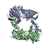 7nueC 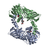 7p2rC 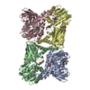 7ppqC 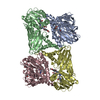 7ppyC 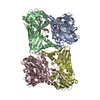 7pu8C 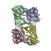 7pucC 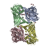 7puqC 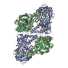 7pv6C 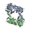 4c03S S: Starting model for refinement C: citing same article ( |
|---|---|
| Similar structure data |
-Links
 PDBj
PDBj
- Assembly
| Deposited unit | 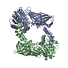
| ||||||||
|---|---|---|---|---|---|---|---|---|---|
| 1 |
| ||||||||
| Unit cell |
|
-Components
| #1: Protein | Mass: 42821.301 Da / Num. of mol.: 2 / Fragment: mouse PRMT6 / Mutation: F315L Source method: isolated from a genetically manipulated source Source: (gene. exp.)  References: UniProt: Q6NZB1, type I protein arginine methyltransferase #2: Chemical | 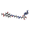  Mass: 608.606 Da / Num. of mol.: 2 Mass: 608.606 Da / Num. of mol.: 2Source method: isolated from a genetically manipulated source Formula: C27H32N10O7 / Feature type: SUBJECT OF INVESTIGATION #3: Water | ChemComp-HOH / |  Mass: 18.015 Da / Num. of mol.: 139 / Source method: isolated from a natural source / Formula: H2O Mass: 18.015 Da / Num. of mol.: 139 / Source method: isolated from a natural source / Formula: H2OHas ligand of interest | Y | |
|---|
-Experimental details
-Experiment
| Experiment | Method: X-RAY DIFFRACTION / Number of used crystals: 1 |
|---|
- Sample preparation
| Crystal | Density Matthews: 2.35 Å3/Da / Density % sol: 47.65 % / Mosaicity: 0.31 ° |
|---|---|
| Crystal grow | Temperature: 298 K / Method: vapor diffusion / pH: 7.5 Details: PEG 3,350 20%, NaNO3 200 mM, Tris-HCl pH 7.5 100 mM |
-Data collection
| Diffraction | Mean temperature: 100 K / Serial crystal experiment: N |
|---|---|
| Diffraction source | Source: ROTATING ANODE / Type: RIGAKU FR-X / Wavelength: 1.54178 Å |
| Detector | Type: DECTRIS PILATUS 300K / Detector: PIXEL / Date: Mar 30, 2017 |
| Radiation | Protocol: SINGLE WAVELENGTH / Monochromatic (M) / Laue (L): M / Scattering type: x-ray |
| Radiation wavelength | Wavelength: 1.54178 Å / Relative weight: 1 |
| Reflection | Resolution: 1.65→45.08 Å / Num. obs: 81095 / % possible obs: 99.6 % / Redundancy: 18.5 % / Biso Wilson estimate: 21.32 Å2 / CC1/2: 0.999 / Rmerge(I) obs: 0.12 / Rpim(I) all: 0.028 / Rrim(I) all: 0.123 / Net I/σ(I): 14.9 |
| Reflection shell | Resolution: 1.65→1.68 Å / Redundancy: 7.3 % / Rmerge(I) obs: 2.058 / Mean I/σ(I) obs: 1.1 / Num. unique obs: 4071 / CC1/2: 0.479 / Rpim(I) all: 0.802 / Rrim(I) all: 2.215 / % possible all: 99.8 |
-Phasing
| Phasing | Method: molecular replacement |
|---|
- Processing
| Software |
| |||||||||||||||||||||||||||||||||||||||||||||||||||||||||||||||||||||||||||||||||||||||||||||||||||||||||
|---|---|---|---|---|---|---|---|---|---|---|---|---|---|---|---|---|---|---|---|---|---|---|---|---|---|---|---|---|---|---|---|---|---|---|---|---|---|---|---|---|---|---|---|---|---|---|---|---|---|---|---|---|---|---|---|---|---|---|---|---|---|---|---|---|---|---|---|---|---|---|---|---|---|---|---|---|---|---|---|---|---|---|---|---|---|---|---|---|---|---|---|---|---|---|---|---|---|---|---|---|---|---|---|---|---|---|
| Refinement | Method to determine structure: MOLECULAR REPLACEMENT Starting model: 4C03 Resolution: 1.65→39.5 Å / SU ML: 0.21 / Cross valid method: THROUGHOUT / σ(F): 1.34 / Phase error: 24.02 / Stereochemistry target values: ML
| |||||||||||||||||||||||||||||||||||||||||||||||||||||||||||||||||||||||||||||||||||||||||||||||||||||||||
| Solvent computation | Shrinkage radii: 0.9 Å / VDW probe radii: 1.11 Å / Solvent model: FLAT BULK SOLVENT MODEL | |||||||||||||||||||||||||||||||||||||||||||||||||||||||||||||||||||||||||||||||||||||||||||||||||||||||||
| Displacement parameters | Biso max: 132.1 Å2 / Biso mean: 33.7923 Å2 / Biso min: 14.3 Å2 | |||||||||||||||||||||||||||||||||||||||||||||||||||||||||||||||||||||||||||||||||||||||||||||||||||||||||
| Refinement step | Cycle: final / Resolution: 1.65→39.5 Å
| |||||||||||||||||||||||||||||||||||||||||||||||||||||||||||||||||||||||||||||||||||||||||||||||||||||||||
| LS refinement shell | Refine-ID: X-RAY DIFFRACTION / Rfactor Rfree error: 0 / % reflection obs: 100 %
|
