 Movie
Movie Controller

[English] 日本語
 Yorodumi
- PDB-7nie: putative glycerol kinase-like proteins anchored on an array of vo... -
+ Open data

- Basic information
| Entry | Database: PDB / ID: 7nie | ||||||
|---|---|---|---|---|---|---|---|
| Title | putative glycerol kinase-like proteins anchored on an array of voltage dependent anion channels in the outer mitochondrial membrane of pig sperm mitochondria | ||||||
 Components Components |
| ||||||
 Keywords Keywords | MEMBRANE PROTEIN / mitochondria / sperm | ||||||
| Function / homology |  Function and homology information Function and homology informationPINK1-PRKN Mediated Mitophagy / Triglyceride biosynthesis / Ub-specific processing proteases / glycerol-3-phosphate biosynthetic process / mitochondrial outer membrane permeabilization / voltage-gated monoatomic anion channel activity / glycerol kinase / glycerol kinase activity / glycerol metabolic process / ceramide binding ...PINK1-PRKN Mediated Mitophagy / Triglyceride biosynthesis / Ub-specific processing proteases / glycerol-3-phosphate biosynthetic process / mitochondrial outer membrane permeabilization / voltage-gated monoatomic anion channel activity / glycerol kinase / glycerol kinase activity / glycerol metabolic process / ceramide binding / voltage-gated monoatomic ion channel activity / oxysterol binding / phosphatidylcholine binding / glycerol catabolic process / monoatomic anion transport / triglyceride metabolic process / cholesterol binding / lipid transport / porin activity / pore complex / sperm midpiece / mitochondrial outer membrane / mitochondrion / ATP binding / membrane Similarity search - Function | ||||||
| Biological species |  | ||||||
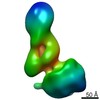
| Method | ELECTRON MICROSCOPY / subtomogram averaging / cryo EM / Resolution: 35 Å | ||||||
 Authors Authors | Leung, M.R. / Zeev-Ben-Mordehai, T. | ||||||
| Funding support |  Netherlands, 1items Netherlands, 1items
| ||||||
 Citation Citation | Journal: Proc Natl Acad Sci U S A / Year: 2021 Title: In-cell structures of conserved supramolecular protein arrays at the mitochondria-cytoskeleton interface in mammalian sperm. Authors: Miguel Ricardo Leung / Riccardo Zenezini Chiozzi / Marc C Roelofs / Johannes F Hevler / Ravi Teja Ravi / Paula Maitan / Min Zhang / Heiko Henning / Elizabeth G Bromfield / Stuart C Howes / ...Authors: Miguel Ricardo Leung / Riccardo Zenezini Chiozzi / Marc C Roelofs / Johannes F Hevler / Ravi Teja Ravi / Paula Maitan / Min Zhang / Heiko Henning / Elizabeth G Bromfield / Stuart C Howes / Bart M Gadella / Albert J R Heck / Tzviya Zeev-Ben-Mordehai /    Abstract: Mitochondria-cytoskeleton interactions modulate cellular physiology by regulating mitochondrial transport, positioning, and immobilization. However, there is very little structural information ...Mitochondria-cytoskeleton interactions modulate cellular physiology by regulating mitochondrial transport, positioning, and immobilization. However, there is very little structural information defining mitochondria-cytoskeleton interfaces in any cell type. Here, we use cryofocused ion beam milling-enabled cryoelectron tomography to image mammalian sperm, where mitochondria wrap around the flagellar cytoskeleton. We find that mitochondria are tethered to their neighbors through intermitochondrial linkers and are anchored to the cytoskeleton through ordered arrays on the outer mitochondrial membrane. We use subtomogram averaging to resolve in-cell structures of these arrays from three mammalian species, revealing they are conserved across species despite variations in mitochondrial dimensions and cristae organization. We find that the arrays consist of boat-shaped particles anchored on a network of membrane pores whose arrangement and dimensions are consistent with voltage-dependent anion channels. Proteomics and in-cell cross-linking mass spectrometry suggest that the conserved arrays are composed of glycerol kinase-like proteins. Ordered supramolecular assemblies may serve to stabilize similar contact sites in other cell types in which mitochondria need to be immobilized in specific subcellular environments, such as in muscles and neurons. | ||||||
| History |
|
- Structure visualization
| Movie |
Movie viewer |
|---|---|
| Structure viewer | Molecule: MolmilJmol/JSmol |
- Downloads & links
-Download
| PDBx/mmCIF format | 7nie.cif.gz | 1.2 MB | Display | PDBx/mmCIF format |
|---|---|---|---|---|
| PDB format | pdb7nie.ent.gz | 1013.7 KB | Display | PDB format |
| PDBx/mmJSON format | 7nie.json.gz | Tree view | PDBx/mmJSON format | |
| Others |  Other downloads Other downloads |
-Validation report
| Arichive directory | https://data.pdbj.org/pub/pdb/validation_reports/ni/7nieftp://data.pdbj.org/pub/pdb/validation_reports/ni/7nie | HTTPS FTP |
|---|
-Related structure data
| Related structure data |  12357MC M: map data used to model this data C: citing same article ( |
|---|---|
| Similar structure data |


-Links
 PDBj
PDBj- Assembly
| Deposited unit | 
|
|---|---|
| 1 |
|
-Components
| #1: Protein | Mass: 53579.547 Da / Num. of mol.: 4 / Source method: isolated from a natural source / Source: (natural) Plasmid details: lamellae prepared by cryo-focused ion beam milling of pig sperm Tissue: sperm / References: UniProt: A0A287BD08, glycerol kinase #2: Protein | Mass: 31637.484 Da / Num. of mol.: 12 / Source method: isolated from a natural source / Source: (natural) Plasmid details: lamellae prepared by cryo-focused ion beam milling of pig sperm Tissue: sperm / References: UniProt: F1S2F6 Has protein modification | Y | |
|---|
-Experimental details
-Experiment
| Experiment | Method: ELECTRON MICROSCOPY |
|---|---|
| EM experiment | Aggregation state: CELL / 3D reconstruction method: subtomogram averaging |
- Sample preparation
| Component | Name: subtomogram average of ordered protein arrays on the axoneme-facing surface of the outer mitochondrial membrane in pig sperm mitochondria (alignment focused on one unit) Type: CELL / Entity ID: all / Source: NATURAL |
|---|---|
| Source (natural) | Organism: |
| Buffer solution | pH: 7.4 |
| Specimen | Embedding applied: NO / Shadowing applied: NO / Staining applied: NO / Vitrification applied: YES |
| Specimen support | Grid material: COPPER / Grid mesh size: 200 divisions/in. / Grid type: Quantifoil |
| Vitrification | Instrument: HOMEMADE PLUNGER / Cryogen name: ETHANE-PROPANE |
- Electron microscopy imaging
| Experimental equipment |  Model: Talos Arctica / Image courtesy: FEI Company |
|---|---|
| Microscopy | Model: FEI TALOS ARCTICA |
| Electron gun | Electron source:  FIELD EMISSION GUN / Accelerating voltage: 200 kV / Illumination mode: FLOOD BEAM FIELD EMISSION GUN / Accelerating voltage: 200 kV / Illumination mode: FLOOD BEAM |
| Electron lens | Mode: BRIGHT FIELD / Alignment procedure: COMA FREE |
| Specimen holder | Cryogen: NITROGEN |
| Image recording | Electron dose: 1.3 e/Å2 / Detector mode: COUNTING / Film or detector model: GATAN K2 SUMMIT (4k x 4k) |
| EM imaging optics | Energyfilter slit width: 20 eV |
- Processing
| EM software |
| ||||||||||||||||||||||||||||
|---|---|---|---|---|---|---|---|---|---|---|---|---|---|---|---|---|---|---|---|---|---|---|---|---|---|---|---|---|---|
| CTF correction | Type: PHASE FLIPPING ONLY | ||||||||||||||||||||||||||||
| Symmetry | Point symmetry: C2 (2 fold cyclic) | ||||||||||||||||||||||||||||
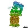
| 3D reconstruction | Resolution: 35 Å / Resolution method: FSC 0.5 CUT-OFF / Num. of particles: 536 / Num. of class averages: 1 / Symmetry type: POINT | ||||||||||||||||||||||||||||
| EM volume selection | Num. of tomograms: 3 / Num. of volumes extracted: 268 / Reference model: random particle from the dataset | ||||||||||||||||||||||||||||
| Atomic model building | Protocol: RIGID BODY FIT Details: Homology models of pig glycerol kinase and VDAC2 were made using Robetta. Rigid body fitting was done with Chimera/ChimeraX. Putative GK proteins only fit in one orientation, but for VDAC ...Details: Homology models of pig glycerol kinase and VDAC2 were made using Robetta. Rigid body fitting was done with Chimera/ChimeraX. Putative GK proteins only fit in one orientation, but for VDAC proteins the rotation around the pore axis remains ambiguous. |
