Netherlands Organisation for Scientific Research (NWO)
740.018.007
Netherlands
Citation
Journal: Proc Natl Acad Sci U S A / Year: 2021 Title: In-cell structures of conserved supramolecular protein arrays at the mitochondria-cytoskeleton interface in mammalian sperm. Authors: Miguel Ricardo Leung / Riccardo Zenezini Chiozzi / Marc C Roelofs / Johannes F Hevler / Ravi Teja Ravi / Paula Maitan / Min Zhang / Heiko Henning / Elizabeth G Bromfield / Stuart C Howes / ...Authors: Miguel Ricardo Leung / Riccardo Zenezini Chiozzi / Marc C Roelofs / Johannes F Hevler / Ravi Teja Ravi / Paula Maitan / Min Zhang / Heiko Henning / Elizabeth G Bromfield / Stuart C Howes / Bart M Gadella / Albert J R Heck / Tzviya Zeev-Ben-Mordehai / Abstract: Mitochondria-cytoskeleton interactions modulate cellular physiology by regulating mitochondrial transport, positioning, and immobilization. However, there is very little structural information ...Mitochondria-cytoskeleton interactions modulate cellular physiology by regulating mitochondrial transport, positioning, and immobilization. However, there is very little structural information defining mitochondria-cytoskeleton interfaces in any cell type. Here, we use cryofocused ion beam milling-enabled cryoelectron tomography to image mammalian sperm, where mitochondria wrap around the flagellar cytoskeleton. We find that mitochondria are tethered to their neighbors through intermitochondrial linkers and are anchored to the cytoskeleton through ordered arrays on the outer mitochondrial membrane. We use subtomogram averaging to resolve in-cell structures of these arrays from three mammalian species, revealing they are conserved across species despite variations in mitochondrial dimensions and cristae organization. We find that the arrays consist of boat-shaped particles anchored on a network of membrane pores whose arrangement and dimensions are consistent with voltage-dependent anion channels. Proteomics and in-cell cross-linking mass spectrometry suggest that the conserved arrays are composed of glycerol kinase-like proteins. Ordered supramolecular assemblies may serve to stabilize similar contact sites in other cell types in which mitochondria need to be immobilized in specific subcellular environments, such as in muscles and neurons.
History
Deposition
Feb 12, 2021
-
Header (metadata) release
Oct 13, 2021
-
Map release
Oct 13, 2021
-
Update
Nov 17, 2021
-
Current status
Nov 17, 2021
Processing site: PDBe / Status: Released
-
Structure visualization
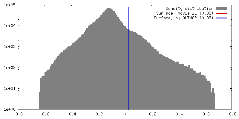
Movie
Surface view with section colored by density value
Entire : ordered protein arrays on the axoneme-facing surface of the outer...
Entire
Name: ordered protein arrays on the axoneme-facing surface of the outer mitochondrial membrane in pig sperm mitochondria
Components
Cell: ordered protein arrays on the axoneme-facing surface of the outer mitochondrial membrane in pig sperm mitochondria
-
Supramolecule #1: ordered protein arrays on the axoneme-facing surface of the outer...
Supramolecule
Name: ordered protein arrays on the axoneme-facing surface of the outer mitochondrial membrane in pig sperm mitochondria type: cell / ID: 1 / Parent: 0
Source (natural)
Organism: Sus scrofa (pig) / Tissue: sperm
-
Experimental details
-
Structure determination
Method
cryo EM
Processing
subtomogram averaging
Aggregation state
cell
-
Sample preparation
Buffer
pH: 7.4
Grid
Model: Quantifoil / Material: COPPER / Mesh: 200 / Support film - Material: CARBON / Support film - topology: HOLEY / Pretreatment - Type: GLOW DISCHARGE
In the structure databanks used in Yorodumi, some data are registered as the other names, "COVID-19 virus" and "2019-nCoV". Here are the details of the virus and the list of structure data.
Jan 31, 2019. EMDB accession codes are about to change! (news from PDBe EMDB page)
EMDB accession codes are about to change! (news from PDBe EMDB page)
The allocation of 4 digits for EMDB accession codes will soon come to an end. Whilst these codes will remain in use, new EMDB accession codes will include an additional digit and will expand incrementally as the available range of codes is exhausted. The current 4-digit format prefixed with “EMD-” (i.e. EMD-XXXX) will advance to a 5-digit format (i.e. EMD-XXXXX), and so on. It is currently estimated that the 4-digit codes will be depleted around Spring 2019, at which point the 5-digit format will come into force.
The EM Navigator/Yorodumi systems omit the EMD- prefix.
Related info.:Q: What is EMD? / ID/Accession-code notation in Yorodumi/EM Navigator
Yorodumi is a browser for structure data from EMDB, PDB, SASBDB, etc.
This page is also the successor to EM Navigator detail page, and also detail information page/front-end page for Omokage search.
The word "yorodu" (or yorozu) is an old Japanese word meaning "ten thousand". "mi" (miru) is to see.
Related info.:EMDB / PDB / SASBDB / Comparison of 3 databanks / Yorodumi Search / Aug 31, 2016. New EM Navigator & Yorodumi / Yorodumi Papers / Jmol/JSmol / Function and homology information / Changes in new EM Navigator and Yorodumi
 Movie
Movie Controller

 Yorodumi
 Open data
 Basic information
 Map data
 Sample
 Authors
 Netherlands, 1 items
  Citation


 Structure visualization
 Movie viewer
 Downloads & links
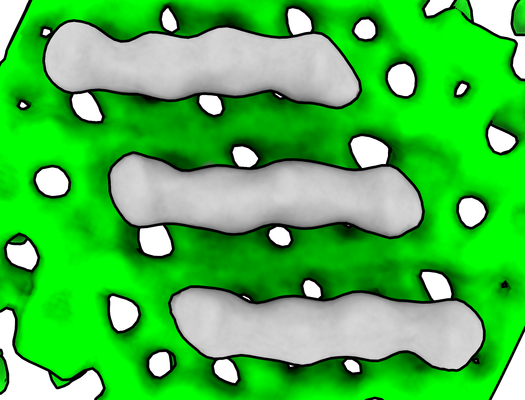 emd_12356.png
 http://ftp.pdbj.org/pub/emdb/structures/EMD-12356


 Z (Sec.)
 Y (Row.)
 X (Col.)


 Sample components
 Processing
 Electron microscopy
 FIELD EMISSION GUN
