Movie
Movie Controller


+ Open data

- Basic information
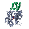
| Entry | Database: PDB / ID: 3byh | ||||||
|---|---|---|---|---|---|---|---|
| Title | Model of actin-fimbrin ABD2 complex | ||||||
 Components Components |
| ||||||
 Keywords Keywords | STRUCTURAL PROTEIN / helical filament / protein polymer / Acetylation / ATP-binding / Cytoplasm / Cytoskeleton / Methylation / Nucleotide-binding / Phosphoprotein | ||||||
| Function / homology |  Function and homology information Function and homology informationpositive regulation of norepinephrine uptake / cellular response to cytochalasin B / Formation of the embryonic stem cell BAF (esBAF) complex / bBAF complex / npBAF complex / brahma complex / nBAF complex / Formation of the canonical BAF (cBAF) complex / regulation of transepithelial transport / Formation of neuronal progenitor and neuronal BAF (npBAF and nBAF) ...positive regulation of norepinephrine uptake / cellular response to cytochalasin B / Formation of the embryonic stem cell BAF (esBAF) complex / bBAF complex / npBAF complex / brahma complex / nBAF complex / Formation of the canonical BAF (cBAF) complex / regulation of transepithelial transport / Formation of neuronal progenitor and neuronal BAF (npBAF and nBAF) / morphogenesis of a polarized epithelium / Formation of the polybromo-BAF (pBAF) complex / structural constituent of postsynaptic actin cytoskeleton / Formation of annular gap junctions / Formation of the dystrophin-glycoprotein complex (DGC) / Gap junction degradation / Formation of the non-canonical BAF (ncBAF) complex / GBAF complex / protein localization to adherens junction / regulation of G0 to G1 transition / Cell-extracellular matrix interactions / dense body / Folding of actin by CCT/TriC / Tat protein binding / postsynaptic actin cytoskeleton / RSC-type complex / Regulation of CDH1 Function / regulation of double-strand break repair / Prefoldin mediated transfer of substrate to CCT/TriC / regulation of nucleotide-excision repair / Adherens junctions interactions / adherens junction assembly / RHOF GTPase cycle / apical protein localization / Sensory processing of sound by outer hair cells of the cochlea / SWI/SNF complex / regulation of mitotic metaphase/anaphase transition / tight junction / Sensory processing of sound by inner hair cells of the cochlea / Interaction between L1 and Ankyrins / positive regulation of T cell differentiation / apical junction complex / positive regulation of double-strand break repair / maintenance of blood-brain barrier / regulation of norepinephrine uptake / positive regulation of stem cell population maintenance / transporter regulator activity / NuA4 histone acetyltransferase complex / Recycling pathway of L1 / Regulation of MITF-M-dependent genes involved in pigmentation / cortical cytoskeleton / establishment or maintenance of cell polarity / nitric-oxide synthase binding / brush border / regulation of G1/S transition of mitotic cell cycle / EPH-ephrin mediated repulsion of cells / negative regulation of cell differentiation / regulation of synaptic vesicle endocytosis / positive regulation of myoblast differentiation / RHO GTPases Activate WASPs and WAVEs / kinesin binding / regulation of protein localization to plasma membrane / RHO GTPases activate IQGAPs / positive regulation of double-strand break repair via homologous recombination / EPHB-mediated forward signaling / cytoskeleton organization / axonogenesis / substantia nigra development / calyx of Held / nitric-oxide synthase regulator activity / FCGR3A-mediated phagocytosis / actin filament / Translocation of SLC2A4 (GLUT4) to the plasma membrane / adherens junction / positive regulation of cell differentiation / cell motility / Regulation of endogenous retroelements by Piwi-interacting RNAs (piRNAs) / RHO GTPases Activate Formins / Signaling by high-kinase activity BRAF mutants / MAP2K and MAPK activation / Regulation of actin dynamics for phagocytic cup formation / structural constituent of cytoskeleton / kinetochore / B-WICH complex positively regulates rRNA expression / VEGFA-VEGFR2 Pathway / platelet aggregation / DNA Damage Recognition in GG-NER / Hydrolases; Acting on acid anhydrides; Acting on acid anhydrides to facilitate cellular and subcellular movement / tau protein binding / Schaffer collateral - CA1 synapse / nuclear matrix / Signaling by RAF1 mutants / cytoplasmic ribonucleoprotein granule / Signaling by moderate kinase activity BRAF mutants / Paradoxical activation of RAF signaling by kinase inactive BRAF / Signaling downstream of RAS mutants / cell-cell junction / Signaling by BRAF and RAF1 fusions / UCH proteinases / nucleosome Similarity search - Function | ||||||
| Biological species |  Homo sapiens (human) Homo sapiens (human) | ||||||
| Method | ELECTRON MICROSCOPY / helical reconstruction / cryo EM / Resolution: 12 Å | ||||||
 Authors Authors | Galkin, V.E. / Orlova, A. / Cherepanova, O. / Lebart, M.C. / Egelman, E.H. | ||||||
 Citation Citation | Journal: Proc Natl Acad Sci U S A / Year: 2008 Title: High-resolution cryo-EM structure of the F-actin-fimbrin/plastin ABD2 complex. Authors: Vitold E Galkin / Albina Orlova / Olga Cherepanova / Marie-Christine Lebart / Edward H Egelman /  Abstract: Many actin binding proteins have a modular architecture, and calponin-homology (CH) domains are one such structurally conserved module found in numerous proteins that interact with F-actin. The ...Many actin binding proteins have a modular architecture, and calponin-homology (CH) domains are one such structurally conserved module found in numerous proteins that interact with F-actin. The manner in which CH-domains bind F-actin has been controversial. Using cryo-EM and a single-particle approach to helical reconstruction, we have generated 12-A-resolution maps of F-actin alone and F-actin decorated with a fragment of human fimbrin (L-plastin) containing tandem CH-domains. The high resolution allows an unambiguous fit of the crystal structure of fimbrin into the map. The interaction between fimbrin ABD2 (actin binding domain 2) and F-actin is different from any interaction previously observed or proposed for tandem CH-domain proteins, showing that the structural conservation of the CH-domains does not lead to a conserved mode of interaction with F-actin. Both the stapling of adjacent actin protomers and the additional closure of the nucleotide binding cleft in F-actin when the fimbrin fragment binds may explain how fimbrin can stabilize actin filaments. A mechanism is proposed where ABD1 of fimbrin becomes activated for binding a second actin filament after ABD2 is bound to a first filament, and this can explain how mutations of residues buried in the interface between ABD2 and ABD1 can rescue temperature-sensitive defects in actin. | ||||||
| History |
|
- Structure visualization
| Movie |
Movie viewer |
|---|---|
| Structure viewer | Molecule: MolmilJmol/JSmol |
- Downloads & links
-Download
| PDBx/mmCIF format | 3byh.cif.gz | 119.3 KB | Display | PDBx/mmCIF format |
|---|---|---|---|---|
| PDB format | pdb3byh.ent.gz | 86.8 KB | Display | PDB format |
| PDBx/mmJSON format | 3byh.json.gz | Tree view | PDBx/mmJSON format | |
| Others |  Other downloads Other downloads |
-Validation report
| Arichive directory | https://data.pdbj.org/pub/pdb/validation_reports/by/3byhftp://data.pdbj.org/pub/pdb/validation_reports/by/3byh | HTTPS FTP |
|---|
-Related structure data
| Similar structure data |
|---|


-Links
 PDBj
PDBj


- Assembly
| Deposited unit | 
|
|---|---|
| 1 | x 10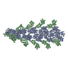
|
| 2 |
|
| 3 | 
|
| Symmetry | Helical symmetry: (Circular symmetry: 1 / Dyad axis: no / N subunits divisor: 1 / Num. of operations: 10 / Rise per n subunits: 27.3 Å / Rotation per n subunits: -166.5 °) |
-Components
| #1: Protein | Mass: 41651.465 Da / Num. of mol.: 1 Source method: isolated from a genetically manipulated source Source: (gene. exp.) Homo sapiens (human) / Gene: PS1TP5BP1 / Production host:  |
|---|---|
| #2: Protein | Mass: 26764.367 Da / Num. of mol.: 1 Source method: isolated from a genetically manipulated source Source: (gene. exp.) Homo sapiens (human) / Production host: |
-Experimental details
-Experiment
| Experiment | Method: ELECTRON MICROSCOPY |
|---|---|
| EM experiment | Aggregation state: FILAMENT / 3D reconstruction method: helical reconstruction |
- Sample preparation
| Component | Name: Actin-Fimbrin ABD2 Complex / Type: COMPLEX Details: helical filament. rotation by -166.5 degrees about the z-axis (x=0, y=0), translation by 27.3 Angstroms along the z-axis |
|---|---|
| Specimen | Embedding applied: NO / Shadowing applied: NO / Staining applied: NO / Vitrification applied: YES |
- Electron microscopy imaging
| Experimental equipment |  Model: Tecnai F20 / Image courtesy: FEI Company |
|---|---|
| Microscopy | Model: FEI TECNAI F20 |
| Electron gun | Electron source:  FIELD EMISSION GUN / Accelerating voltage: 200 kV / Illumination mode: FLOOD BEAM FIELD EMISSION GUN / Accelerating voltage: 200 kV / Illumination mode: FLOOD BEAM |
| Electron lens | Mode: BRIGHT FIELD |
| Image recording | Film or detector model: GENERIC FILM |
| Image scans | Scanner model: NIKON COOLSCAN |
- Processing
| CTF correction | Details: micrographs were multiplied by the CTF function, and the final reconstruction was then divided by the summed squared CTFs | |||||||||||||||||||||
|---|---|---|---|---|---|---|---|---|---|---|---|---|---|---|---|---|---|---|---|---|---|---|
| 3D reconstruction | Method: IHRSR / Resolution: 12 Å / Nominal pixel size: 2.38 Å / Actual pixel size: 2.38 Å / Magnification calibration: TMV / Symmetry type: HELICAL | |||||||||||||||||||||
| Atomic model building |
| |||||||||||||||||||||
| Refinement step | Cycle: LAST
|