 Movie
Movie Controller

[English] 日本語
 Yorodumi
- PDB-7nef: Fucosylated linear peptide Fln65 bound to the fucose binding lect... -
+ Open data

- Basic information
| Entry | Database: PDB / ID: 7nef | ||||||
|---|---|---|---|---|---|---|---|
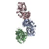
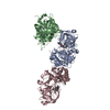
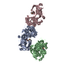
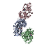
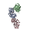
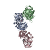
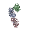
| Title | Fucosylated linear peptide Fln65 bound to the fucose binding lectin LecB PA-IIL from Pseudomonas aeruginosa at 1.5 Angstrom resolution | ||||||
 Components Components |
| ||||||
 Keywords Keywords | ANTIBIOTIC / Antimicrobial peptide / Lectin | ||||||
| Function / homology |  Function and homology information Function and homology informationsingle-species biofilm formation / carbohydrate binding / metal ion binding Similarity search - Function | ||||||
| Biological species |   Pseudomonas aeruginosa (bacteria) Pseudomonas aeruginosa (bacteria)synthetic construct (others) | ||||||
| Method |  X-RAY DIFFRACTION / SYNCHROTRON / MOLECULAR REPLACEMENT / molecular replacement / Resolution: 1.51 Å X-RAY DIFFRACTION / SYNCHROTRON / MOLECULAR REPLACEMENT / molecular replacement / Resolution: 1.51 Å | ||||||
 Authors Authors | Personne, H. / Baeriswyl, S. / Stocker, A. / Reymond, J.-L. | ||||||
| Funding support |  Switzerland, 1items Switzerland, 1items
| ||||||
 Citation Citation | Journal: Rsc Chem Biol / Year: 2021 Title: A mixed chirality alpha-helix in a stapled bicyclic and a linear antimicrobial peptide revealed by X-ray crystallography. Authors: Baeriswyl, S. / Personne, H. / Di Bonaventura, I. / Kohler, T. / van Delden, C. / Stocker, A. / Javor, S. / Reymond, J.L. #1: Journal: Rsc Chem Biol / Year: 2021Title: A mixed chirality alpha-helix in a stapled bicyclic and a linear antimicrobial peptide revealed by X-ray crystallography. Authors: Baeriswyl, S. / Personne, H. / Di Bonaventura, I. / Kohler, T. / van Delden, C. / Stocker, A. / Javor, S. / Reymond, J.L. #2: Journal: Rsc Chem Biol / Year: 2021Title: A mixed chirality alpha-helix in a stapled bicyclic and a linear antimicrobial peptide revealed by X-ray crystallography. Authors: Baeriswyl, S. / Personne, H. / Di Bonaventura, I. / Kohler, T. / van Delden, C. / Stocker, A. / Javor, S. / Reymond, J.L. #3: Journal: Chemrxiv / Year: 2021Title: Mixed chirality alpha-helix in a stapled bicyclic and a linear antimicrobial peptide revealed by X-ray crystallography Authors: Personne, H. / Baeriswyl, S. / Di Bonaventura, I. / Kohler, T. / van Delden, C. / Stocker, A. / Javor, S. / Reymond, J.-L. | ||||||
| History |
|
- Structure visualization
| Structure viewer | Molecule: MolmilJmol/JSmol |
|---|
- Downloads & links
-Download
| PDBx/mmCIF format | 7nef.cif.gz | 552.6 KB | Display | PDBx/mmCIF format |
|---|---|---|---|---|
| PDB format | pdb7nef.ent.gz | 459.1 KB | Display | PDB format |
| PDBx/mmJSON format | 7nef.json.gz | Tree view | PDBx/mmJSON format | |
| Others |  Other downloads Other downloads |
-Validation report
| Arichive directory | https://data.pdbj.org/pub/pdb/validation_reports/ne/7nefftp://data.pdbj.org/pub/pdb/validation_reports/ne/7nef | HTTPS FTP |
|---|
-Related structure data
| Related structure data |  6y0uC  6y0vC 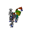 6y13C 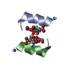 6y14C  6y1sC  7newC 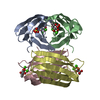 1oxcS S: Starting model for refinement C: citing same article ( |
|---|---|
| Similar structure data |


-Links
 PDBj
PDBj- Assembly
| Deposited unit | 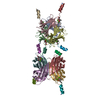
| ||||||||
|---|---|---|---|---|---|---|---|---|---|
| 1 |
| ||||||||
| Unit cell |
|
-Components
| #1: Protein | Mass: 11865.905 Da / Num. of mol.: 8 Source method: isolated from a genetically manipulated source Details: Fucose-binding Lectin LecB PA-IIL from Pseudomonas aeruginosa Source: (gene. exp.) Pseudomonas aeruginosa (bacteria)Gene: lecB, C0044_25260, CAZ10_21840, DT376_00595, DY979_15445, ECC04_10105, EFK27_13700, EGV95_09240, EGY23_15550, IPC669_23070, PA5486_01888, PAERUG_E15_London_28_01_14_00983, PAMH19_1713, RW109_RW109_02453 Production host: #2: Protein/peptide | Mass: 1324.847 Da / Num. of mol.: 8 / Source method: obtained synthetically / Details: Fucosylated linear peptide Fln65 / Source: (synth.) synthetic construct (others) #3: Chemical | ChemComp-CA /   Mass: 40.078 Da / Num. of mol.: 16 / Source method: obtained synthetically / Formula: Ca Mass: 40.078 Da / Num. of mol.: 16 / Source method: obtained synthetically / Formula: Ca#4: Sugar | ChemComp-ZDC / 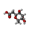  Type: D-saccharide / Mass: 206.193 Da / Num. of mol.: 8 Type: D-saccharide / Mass: 206.193 Da / Num. of mol.: 8Source method: isolated from a genetically manipulated source Formula: C8H14O6 #5: Water | ChemComp-HOH / |  Mass: 18.015 Da / Num. of mol.: 1415 / Source method: isolated from a natural source / Formula: H2O Mass: 18.015 Da / Num. of mol.: 1415 / Source method: isolated from a natural source / Formula: H2OHas ligand of interest | N | Has protein modification | Y | |
|---|
-Experimental details
-Experiment
| Experiment | Method: X-RAY DIFFRACTION / Number of used crystals: 1 |
|---|
- Sample preparation
| Crystal | Density Matthews: 2.69 Å3/Da / Density % sol: 54.24 % |
|---|---|
| Crystal grow | Temperature: 291 K / Method: vapor diffusion, sitting drop / Details: 0.2M Magnesium formate dihydrate |
-Data collection
| Diffraction | Mean temperature: 100 K / Serial crystal experiment: N |
|---|---|
| Diffraction source | Source: SYNCHROTRON / Site: SLS / Beamline: X06DA / Wavelength: 0.976 Å |
| Detector | Type: DECTRIS PILATUS 2M-F / Detector: PIXEL / Date: Nov 2, 2020 |
| Radiation | Protocol: MAD / Monochromatic (M) / Laue (L): M / Scattering type: x-ray |
| Radiation wavelength | Wavelength: 0.976 Å / Relative weight: 1 |
| Reflection | Resolution: 1.507→48.591 Å / Num. obs: 336482 / % possible obs: 96.9 % / Redundancy: 3.18 % / CC1/2: 0.997 / Rrim(I) all: 0.117 / Net I/σ(I): 8.23 |
| Reflection shell | Resolution: 1.51→1.6 Å / Redundancy: 2.36 % / Mean I/σ(I) obs: 0.12 / Num. unique obs: 50384 / CC1/2: 0.685 / Rrim(I) all: 0.933 / % possible all: 89.8 |
-Phasing
| Phasing | Method: molecular replacement |
|---|
- Processing
| Software |
| |||||||||||||||||||||||||||||||||||||||||||||||||||||||||||||||||||||||||||||||||||||||||||||||||||||||||||||||||||||||||||||||||||||||||||||||||||||||||||||||||||||||||||||||||||||||||||||||||||||||||||||||||||||||||
|---|---|---|---|---|---|---|---|---|---|---|---|---|---|---|---|---|---|---|---|---|---|---|---|---|---|---|---|---|---|---|---|---|---|---|---|---|---|---|---|---|---|---|---|---|---|---|---|---|---|---|---|---|---|---|---|---|---|---|---|---|---|---|---|---|---|---|---|---|---|---|---|---|---|---|---|---|---|---|---|---|---|---|---|---|---|---|---|---|---|---|---|---|---|---|---|---|---|---|---|---|---|---|---|---|---|---|---|---|---|---|---|---|---|---|---|---|---|---|---|---|---|---|---|---|---|---|---|---|---|---|---|---|---|---|---|---|---|---|---|---|---|---|---|---|---|---|---|---|---|---|---|---|---|---|---|---|---|---|---|---|---|---|---|---|---|---|---|---|---|---|---|---|---|---|---|---|---|---|---|---|---|---|---|---|---|---|---|---|---|---|---|---|---|---|---|---|---|---|---|---|---|---|---|---|---|---|---|---|---|---|---|---|---|---|---|---|---|---|
| Refinement | Method to determine structure: MOLECULAR REPLACEMENT Starting model: 1OXC Resolution: 1.51→48.59 Å / SU ML: 0.18 / Cross valid method: THROUGHOUT / σ(F): 0.9 / Phase error: 21.04 / Stereochemistry target values: ML
| |||||||||||||||||||||||||||||||||||||||||||||||||||||||||||||||||||||||||||||||||||||||||||||||||||||||||||||||||||||||||||||||||||||||||||||||||||||||||||||||||||||||||||||||||||||||||||||||||||||||||||||||||||||||||
| Solvent computation | Shrinkage radii: 0.9 Å / VDW probe radii: 1.11 Å / Solvent model: FLAT BULK SOLVENT MODEL | |||||||||||||||||||||||||||||||||||||||||||||||||||||||||||||||||||||||||||||||||||||||||||||||||||||||||||||||||||||||||||||||||||||||||||||||||||||||||||||||||||||||||||||||||||||||||||||||||||||||||||||||||||||||||
| Displacement parameters | Biso max: 95.67 Å2 / Biso mean: 21.0708 Å2 / Biso min: 9.11 Å2 | |||||||||||||||||||||||||||||||||||||||||||||||||||||||||||||||||||||||||||||||||||||||||||||||||||||||||||||||||||||||||||||||||||||||||||||||||||||||||||||||||||||||||||||||||||||||||||||||||||||||||||||||||||||||||
| Refinement step | Cycle: final / Resolution: 1.51→48.59 Å
| |||||||||||||||||||||||||||||||||||||||||||||||||||||||||||||||||||||||||||||||||||||||||||||||||||||||||||||||||||||||||||||||||||||||||||||||||||||||||||||||||||||||||||||||||||||||||||||||||||||||||||||||||||||||||
| LS refinement shell | Refine-ID: X-RAY DIFFRACTION / Rfactor Rfree error: 0 / Total num. of bins used: 30
| |||||||||||||||||||||||||||||||||||||||||||||||||||||||||||||||||||||||||||||||||||||||||||||||||||||||||||||||||||||||||||||||||||||||||||||||||||||||||||||||||||||||||||||||||||||||||||||||||||||||||||||||||||||||||
| Refinement TLS params. | Method: refined / Origin x: 21.181 Å / Origin y: -7.0985 Å / Origin z: -29.1351 Å
| |||||||||||||||||||||||||||||||||||||||||||||||||||||||||||||||||||||||||||||||||||||||||||||||||||||||||||||||||||||||||||||||||||||||||||||||||||||||||||||||||||||||||||||||||||||||||||||||||||||||||||||||||||||||||
| Refinement TLS group |
|
